. 2017 Feb 17;292(7):2670-2678.
doi: 10.1074/jbc.M116.770016. Epub 2016 Dec 28.

Cell-based Fluorescence Complementation Reveals a Role for HIV-1 Nef Protein Dimerization in AP-2 Adaptor Recruitment and CD4 Co-receptor Down-regulation

Affiliations

Cell-based Fluorescence Complementation Reveals a Role for HIV-1 Nef Protein Dimerization in AP-2 Adaptor Recruitment and CD4 Co-receptor Down-regulation

Sherry T Shu et al. J Biol Chem. .

Abstract

The HIV-1 Nef accessory factor enhances viral infectivity, immune evasion, and AIDS progression. Nef triggers rapid down-regulation of CD4 via the endocytic adaptor protein 2 (AP-2) complex, a process linked to enhanced viral infectivity and immune escape. Here, we describe a bimolecular fluorescence complementation (BiFC) assay to visualize the interaction of Nef with AP-2 and CD4 in living cells. Interacting protein pairs were fused to complementary non-fluorescent fragments of YFP and co-expressed in 293T cells. Nef interactions with both CD4 and AP-2 resulted in complementation of YFP and a bright fluorescent signal by confocal microcopy that localized to the cell periphery. Co-expression of the AP-2 α subunit enhanced the Nef·AP-2 σ2 subunit BiFC signal and vice versa, suggesting that the AP-2 α-σ2 hemicomplex interacts cooperatively with Nef. Mutagenesis of Nef amino acids Arg-134, Glu-174, and Asp-175, which stabilize Nef for AP-2 α-σ2 binding in a recent co-crystal structure, substantially reduced AP-2 interaction without affecting CD4 binding. A dimerization-defective mutant of Nef failed to interact with either CD4 or AP-2 in the BiFC assay, indicating that Nef quaternary structure is required for CD4 and AP-2 recruitment as well as CD4 down-regulation. A small molecule previously shown to bind the Nef dimerization interface also reduced Nef interactions with AP-2 and CD4 and restored CD4 expression to the surface of HIV-infected cells. Our findings provide a mechanistic explanation for previous observations that dimerization-defective Nef mutants fail to down-regulate CD4 and validate the Nef dimerization interface as a target site for antiretroviral drug development.

Keywords: AP-2; Nef; bimolecular fluorescence complementation; cluster of differentiation 4 (CD4); dimerization; endocytosis; human immunodeficiency virus (HIV); protein-protein interaction.


Conflict of interest statement

The authors declare that they have no conflicts of interest with the contents of this article

Figures
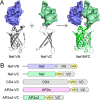
FIGURE 1.
FIGURE 1.
BiFC principle and expression constructs used in this study. A, illustration of the BiFC principle using crystal structures of the Nef homodimer (PDB code 1EFN) and the reassembled Venus variant of YFP (PDB code 3AKO). Non-fluorescent N-terminal (VN) or C-terminal (VC) fragments of Venus are fused to the C terminus of Nef and co-expressed in the same cell. Nef homodimerization brings the Venus fragments into close proximity, resulting in functional complementation and fluorescence (Nef-BiFC, right). B, non-fluorescent Venus fragments were also fused to the C termini of CD4 and the AP-2 α and σ2 subunits. HA and V5 epitope tags were included to enable immunofluorescent staining. The Nef BiFC expression vectors are constructed so that the N-terminal myristoylation signal sequence is intact for membrane targeting.
FIGURE 2.
FIGURE 2.
Visualization of Nef interactions with CD4 and AP-2 subunits by BiFC. Nef-VN was co-expressed with CD4-VC, AP-2 α-VC, and AP-2 σ2-VC in 293T cells, and BiFC was assessed by confocal microscopy (green, left column). Nef expression was detected by immunostaining with anti-HA antibodies (red, center column). Expression of CD4, AP-2 α, and AP-2 σ2 was detected by immunostaining with anti-V5 antibodies (blue, left column).
FIGURE 3.
FIGURE 3.
Subcellular localization of Nef interactions with CD4 and AP-2 subunits by BiFC. Nef-VN was co-expressed with CD4-VC, AP-2 α-VC, and AP-2 σ2-VC in 293T cells, and BiFC was assessed by confocal microscopy (green). As a negative control, an Itk BiFC construct consisting of the pleckstrin homology and SH3 domains fused to VN (Itk-VN) (37) was co-expressed with AP-2 α-VC. Nef expression was detected by immunostaining with anti-HA antibodies (red), whereas expression of CD4, AP-2 α, AP-2 σ2, and Itk was detected by immunostaining with anti-V5 or Itk antibodies (blue). Merged images are shown in the right-hand column.
FIGURE 4.
FIGURE 4.
Nef and AP-2 BiFC fusion proteins retain CD4 down-regulation function. Left panel, CD4 (without VC fusion) was expressed alone in 293T cells or together with unfused versions of the AP-2 α and σ2 subunits, Nef-YFP, or all three proteins combined. Cell-surface CD4 expression was determined by flow cytometry using an APC-conjugated CD4 antibody. Each histogram shows the cell counts versus fluorescence intensity of APC-CD4 in each cell population; ∼10,000 cells were analyzed for each condition. No fluorescence was observed in untransfected 293T cells (not shown). This experiment was repeated in the same way using the corresponding BiFC fusion constructs shown in Fig. 1 (right panel).
FIGURE 5.
FIGURE 5.
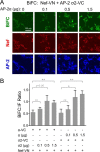
AP-2 α and σ2 subunits interact cooperatively with Nef. A, Nef-VN and AP-2 σ2-VC were expressed in 293T cells either alone or in the presence of increasing amounts of the unfused AP-2 α subunit (microgram amounts of the AP-2 α expression vector are indicated above the image panels). Nef interaction with AP-2 σ2 was assessed as the BiFC signal by confocal microscopy (green, top row). Nef and AP-2 subunit expression were confirmed by immunostaining with anti-HA antibodies (red, 2nd row) and anti-V5 antibodies (blue, bottom row), respectively. B, image analysis of Nef interaction with AP-2 σ2 by BiFC. Fluorescence intensities for the BiFC and Nef immunofluorescence channels were determined for a minimum of 100 cells from A using ImageJ, and BiFC to IF signal ratios were calculated for each condition. The experiment was repeated in triplicate, and the results are presented as the mean ratios ± S.D. Also shown are the results from a complementary experiment with the Nef-VN plus AP-2 α-VC BiFC pair and titration of unfused AP-2 σ2. Unpaired Student's t tests were performed among the groups shown; *, p < 0.05; **, p < 0.001; ns, not significant.
FIGURE 6.
FIGURE 6.
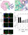
Stabilizing contacts in the Nef central loop are required for interaction with the AP-2 α-σ2 hemicomplex. A, molecular model of the X-ray crystal structure of the HIV-1 Nef core in complex with the AP-2 α-σ2 hemicomplex. Left, Nef core is rendered in purple, with the central loop highlighted in cyan. This Nef loop interacts with both the α (pink) and σ2 (green) subunits, with most of the contact surface involving σ2. Right, Nef dileucine motif within the central loop (Leu-164/Leu-165) makes direct contact with the AP-2 σ2 subunit. A salt bridge between Nef central loop residue Asp-175 and Arg-134 in the Nef core stabilizes the loop conformation for AP-2 binding. A second Nef core-to-loop hydrogen bond (Asp-174 with Gln-104) may also contribute to stabilization. (Note that in the SF2 Nef clone used in our studies, amino acid position 174 is replaced with glutamate instead of aspartate.) Models were produced using PyMOL and PDB code 4NEE (25). B, BiFC assays of Nef central loop mutants with AP-2 subunits. VN fusion proteins of wild type (WT) Nef, along with the central loop mutants R134E and E174A/D175A (ED-AA) were co-expressed with AP-2 α-VC and σ2-VC in 293T cells. Cells were counterstained with antibodies to the HA tag on Nef and the V5 tag on the partner proteins, followed by confocal imaging for BiFC (green), partner protein expression (blue, insets), and Nef expression (red, insets). C, image analysis of Nef interaction with AP-2 α and σ2 subunits by BiFC. Fluorescence intensities for the BiFC and Nef IF channels were determined for a minimum of 100 cells using ImageJ, and BiFC to IF signal intensity ratios were calculated. The experiment was repeated in triplicate, and the results are presented as the mean ratios ± S.D. Unpaired Student's t tests comparing control values from lane 1 yielded the following results: lane 1 versus 3 and lane 1 versus 4, p < 0.05; lane 1 versus 5, p = 0.056; lane 1 versus 6, p = 0.093. D, mutation of the Nef internal loop does not impact CD4 recruitment. VN fusion proteins of the mutant forms of Nef from B were co-expressed with CD4-VC in 293T cells, followed by counterstaining for Nef and V5, and confocal imaging for BiFC (green), partner protein expression (blue, insets), and Nef expression (red, insets).
FIGURE 7.
FIGURE 7.
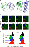
Dimerization-defective mutant of Nef shows reduced AP-2 and CD4 interaction and no longer down-regulates CD4. A, molecular model of the Nef dimer based on the crystal structure of Nef in complex with an Src family kinase SH3 domain (PDB code 1EFN) (30). Left, two Nef monomers are colored green and purple, and the dimer interface is formed by the orthogonal juxtaposition of their αB helices. Right, close-up view of the helical interface, showing the side chains of four hydrophobic residues that pack together to stabilize this dimer (Ile-109, Leu-112, Tyr-115, and Phe-121). Replacement of these four residues with aspartate (Nef-4D mutant) results in a charged surface that prevents dimerization but retains the helical structure (33). B, BiFC assays of the Nef-4D mutant with AP-2 subunits and CD4. VN fusion proteins of the wild type (WT) and 4D forms of Nef were co-expressed with AP-2 α-VC, σ2-VC, and CD4-VC in 293T cells. Cells were counterstained with antibodies to the HA tag on Nef and the V5 tag on the partner proteins, followed by confocal imaging for BiFC (green), partner protein expression (blue, insets), and Nef expression (red, insets). C, flow cytometry for cell-surface CD4. Left, CD4 was expressed alone in 293T cells or together with the wild-type, R134E, or 4D forms of Nef. Cell surface CD4 expression was determined by flow cytometry using an APC-conjugated CD4 antibody. Each histogram shows the cell counts versus fluorescence intensity of APC-CD4 in each cell population; ∼10,000 cells were analyzed for each condition. No fluorescence was observed in untransfected 293T cells (not shown). Right, CD4 down-regulation experiment was performed as before except the AP-2 α and σ2 subunit expression plasmids were added to the co-transfection.
FIGURE 8.
FIGURE 8.
Pharmacological inhibition of Nef dimerization reduces AP-2 recruitment and rescues CD4 down-regulation in HIV-infected cells. A, diphenylpyrazolodiazene Nef inhibitor B9 (structure at right) is predicted to bind to the Nef dimerization interface. The two Nef monomers are modeled in green and purple. The most energetically favorable docking site for B9 was previously identified at the dimer interface using AutoDock Vina (36). Carbon atoms of the B9 scaffold are rendered in yellow. B, B9 treatment reduces Nef dimerization and effector protein recruitment in the BiFC assay. The wild type Nef-VN fusion protein was co-expressed with Nef-VC, AP-2 α-VC, AP-2 σ2-VC, and CD4-VC in 293T cells in the presence or absence of B9 (3 μm). Cells were also transfected with Nef-VC plus Itk-VN in the presence of B9 as a control. Cells were then counterstained with antibodies to the HA tag on Nef, followed by confocal imaging for BiFC and Nef expression. Fluorescence intensity for each protein-protein interaction (BiFC) and Nef expression (Nef-IF) was determined as described in the legends to Figs. 5 and 6, and the BiFC/Nef-IF ratios were calculated. Mean ratios from the B9-treated cultures are presented as percent of the corresponding untreated controls from three independent experiments ± S.D. C, B9 treatment restores cell surface CD4 expression in HIV-infected cells. CEM-GFP cells were infected with wild-type HIV-1 (WT), a Nef-defective mutant (ΔNef), or with WT in the presence of B9 (3 μm). Surface expression of CD4 was determined by flow cytometry as described under “Experimental Procedures.” Data were normalized to CD4 surface levels in uninfected cells (Con) and are presented as the mean ± S.D. from at least three independent experiments. Unpaired Student's t tests showed significant differences between WT and ΔNef (**, p < 0.005) and WT in the presence versus absence of B9 (*, p < 0.05) with no significant difference between ΔNef and WT + B9 (ns).
FIGURE 9.
FIGURE 9.
Structural alignment of a Nef dimer with the Nef·AP-2 complex. The Nef dimer present in complex with the Hck SH3-SH2 dual domain (PDB code 4U5W) (31) was aligned with the Nef·AP-2 crystal complex (PDB code 4NEE) (25). Left, one Nef monomer from 4U5W was aligned with the Nef core in 4NEE (Nef core in gray). The two Nef cores adopt nearly identical folds, with the exception of the structured central loop (cyan), which is present only in the AP-2 complex. Nef residues implicated in CD4 down-regulation are shown in yellow. The AP-2 α and σ2 subunits are rendered in pink and green, respectively. Right, position of the second Nef monomer (NefB) from the 4U5W dimer is shown in orange. The two α-helices that form the Nef dimer interface are indicated (αBA and αBB).

References

    1. Foster J. L., and Garcia J. V. (2008) HIV-1 Nef: at the crossroads. Retrovirology 5, 84. - PMC - PubMed
    1. Kestler H. W. 3rd, Ringler D. J., Mori K., Panicali D. L., Sehgal P. K., Daniel M. D., and Desrosiers R. C. (1991) Importance of the nef gene for maintenance of high viral loads and for development of AIDS. Cell 65, 651–662 - PubMed
    1. Deacon N. J., Tsykin A., Solomon A., Smith K., Ludford-Menting M., Hooker D. J., McPhee D. A., Greenway A. L., Ellett A., Chatfield C., Lawson V. A., Crowe S., Maerz A., Sonza S., Learmont J., et al. (1995) Genomic structure of an attenuated quasi species of HIV-1 from a blood transfusion donor and recipients. Science 270, 988–991 - PubMed
    1. Kirchhoff F., Greenough T. C., Brettler D. B., Sullivan J. L., and Desrosiers R. C. (1995) Absence of intact nef sequences in a long-term survivor with nonprogressive HIV-1 infection. N. Engl. J. Med. 332, 228–232 - PubMed
    1. Jolicoeur P. (2011) The CD4C/HIV(Nef)transgenic model of AIDS. Curr. HIV Res. 9, 524–530 - PubMed

Publication types

Substances

LinkOut - more resources