. 2017 Feb 10;292(6):2255-2265.
doi: 10.1074/jbc.M116.760330. Epub 2016 Dec 28.

PRMT5 C-terminal Phosphorylation Modulates a 14-3-3/PDZ Interaction Switch

Affiliations

PRMT5 C-terminal Phosphorylation Modulates a 14-3-3/PDZ Interaction Switch

Alexsandra B Espejo et al. J Biol Chem. .

Abstract

PRMT5 is the primary enzyme responsible for the deposition of the symmetric dimethylarginine in mammalian cells. In an effort to understand how PRMT5 is regulated, we identified a threonine phosphorylation site within a C-terminal tail motif, which is targeted by the Akt/serum- and glucocorticoid-inducible kinases. While investigating the function of this posttranslational modification, we serendipitously discovered that its free C-terminal tail binds PDZ domains (when unphosphorylated) and 14-3-3 proteins (when phosphorylated). In essence, a phosphorylation event within the last few residues of the C-terminal tail generates a posttranslational modification-dependent PDZ/14-3-3 interaction "switch." The C-terminal motif of PRMT5 is required for plasma membrane association, and loss of this switching capacity is not compatible with life. This signaling phenomenon was recently reported for the HPV E6 oncoprotein but has not yet been observed for mammalian proteins. To investigate the prevalence of PDZ/14-3-3 switching in signal transduction, we built a protein domain microarray that harbors PDZ domains and 14-3-3 proteins. We have used this microarray to interrogate the C-terminal tails of a small group of candidate proteins and identified ERBB4, PGHS2, and IRK1 (as well as E6 and PRMT5) as conforming to this signaling mode, suggesting that PDZ/14-3-3 switching may be a broad biological paradigm.

Keywords: 14–3-3 protein; PDZ domain; cell signaling; protein arginine N-methyltransferase 5 (PRMT5); protein methylation; protein phosphorylation.


Figures

FIGURE 1.
FIGURE 1.
PRMT5 is phosphorylated at threonine 634 in its C terminus. A, schematic representation of GFP-PRMT5 (amino acids 340–637). The predicted Akt-phosphorylated site (Thr-634) is shown. The signature methyltransferase motifs are boxed. B, HeLa cells were transfected with GFP-PRMT5 (WT or T634A) and Myr-Akt and then treated with 0.1 μm calyculin A. Cells were lysed and subjected to immunoprecipitation (IP) with α-GFP. Immunoprecipitates and input samples were then blotted with pan-phosphothreonine (α-pT) and α-GFP antibodies. C, SK-CO15 cells transiently expressing GFP-PRMT5 (WT or T634A) were treated with angiotensin II, dexamethasone, and forskolin or co-transfected with Myr-Akt. Cell lysates were immunoprecipitated with α-GFP followed by Western blotting with pan-phosphothreonine and α-GFP antibodies. The input samples were blotted with α-Akt. β-Actin was used as a loading control. D, the bar graph represents the normalized ratio of phosphorylated GFP-PRMT5 (pan-phosphothreonine) and total GFP-PRMT5 (α-GFP). Quantification was performed by densitometry of the top panel in C and a duplicate experiment. Three independent measurements were taken for each band from the two different blots. S.D. is denoted by error bars; *, p < 0.01; **, p < 0.001, generated by an unpaired Student's t test.
FIGURE 2.
FIGURE 2.
In vitro screening of kinases that phosphorylate a PRMT5 C-terminal peptide. A, the activity of 295 protein kinases was tested against a PRMT5 C-terminal peptide. These assays were based on the direct quantification of radiolabeled phosphate from ATP (γ-33P) on to the peptide substrate. These assays were performed by Kinexus. In vitro kinase activity was ranked as excellent (>5.5 pmol/min), good (4.6–5.4 pmol/min), low (1.8–4.5 pmol/min), and non-phosphorylated (<1.7 pmol/min). B, the 18 best kinases (excellent and good ranking) were used to validate the initial in vitro phosphorylation screen using PRMT5 WT and T634A C-terminal peptides as substrates.
FIGURE 3.
FIGURE 3.
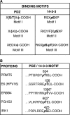
PDZ domain and 14-3-3 share a common binding motif. A, comparison of 14-3-3 versus PDZ domain binding motifs. The consensus binding sequences of the PDZ-binding motif I and 14-3-3-binding motif III are very similar, differing only in the phosphorylation-dependent characteristic of 14-3-3 interactions. Single-letter amino acid codes are used; X, any residue; φ, a hydrophobic residue; P, phosphorylation. B, schematic diagram of proteins harboring overlapping PDZ and 14-3-3 binding motifs, displaying predicted (*) phosphorylation sites (ERBB4 and PGHS2) and reported phosphorylation sites (PRMT5, IRK1, and E6). Predictions were done using iGPS version 1.0 software.
FIGURE 4.
FIGURE 4.
Map of PDZ/14-3-3 protein microarray. The protein microarray, consisting of GST fusion proteins of all 14-3-3 isoforms and 76 PDZ domains. Each GST fusion protein is arrayed in duplicate, at a different angle to facilitate the identification of the spots. GST was used as negative control.
FIGURE 5.
FIGURE 5.
Phosphorylation triggers switching between 14-3-3 and PDZ interactions. A, PRMT5, ERBB4, E6 (HPV16), PGHS2, and IRK1 unphosphorylated and phosphorylated peptides were labeled with Cy5 (red) and Cy3 (green), respectively, and used to probe a protein microarray containing PDZ domains and 14-3-3 GST fusion proteins. The bottom right panel shows the array probed with α-GST for the loading control. The PDZ domains (red) and 14-3-3 proteins (green) are blocked. B, graphical depiction of the interactions observed in A. Red and green squares, interactions with unphosphorylated and phosphorylated peptides, respectively.
FIGURE 6.
FIGURE 6.
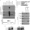
PRMT5 peptide pull-downs confirm an in vitro interaction with NHERF2. A, GST-fused PDZ domains of GRIP1 (residues 672–754), MPP7 (residues 139–220), PDZ-LIM55 (residues 2–85), NHERF1 FL (residues 1–355), NHERF2 FL (residues 1–337), SCRIB (residues 714–801), PDZ-LIM2 (residues 1–84), and GST were incubated with biotinylated PRMT5 C terminus unphosphorylated peptide. Bound proteins were detected with α-GST antibody (short and long exposure are shown). Peptide loading was assessed with HRP-conjugated streptavidin (SA-HRP). The Coomassie stain demonstrates roughly equal input of the GST fusion proteins. B, schematic representation of the constructs used for peptide pull-down in C. C, purified recombinant GST, GST-tagged human NHERF2 full-length (NHERF2-PDZ 1–2), PDZ1 (amino acids 1–152), PDZ2 (amino acids 107–337), and 14-3-3ϵ were incubated with biotinylated PRMT5 C terminus unphosphorylated and Thr-634-phosphorylated peptides bound to streptavidin-agarose beads and detected by α-GST. Left lane, inputs of the GST fusion proteins. D, 293T cells were transfected with constructs expressing GFP-14-3-3ϵ and Myc-PRMT5 wild type or T634A mutant. Cell lysates were then incubated with normal mouse IgG or α-Myc antibody. Immunocomplexes were captured by Protein A beads and detected by either α-Myc or α-GFP. IB, immunoblotting.
FIGURE 7.
FIGURE 7.
PRMT5 interacts with NHERF2 and associates with the plasma membrane. A, control and PRMT5 knockdown (KD) HeLa cells were treated with or without calyculin A, and the lysates were used for GST pull-down assays. 14-3-3ϵ was able to pull down PRMT5 in the calyculin A-treated cells, whereas NHERF2 pulled down PRMT5 from the untreated cell lysate. B, PRMT5 KD HeLa cells were transiently transfected with shRNA-resistant Myc-PRMT5 WT, Δ (deletion of last 6 amino acids), and T634A mutant. Cells were subjected to calyculin A treatment, and pull-down assays were performed as described in A. C, SK-CO15 cells, which express high levels of NHERF2, were fractionated by ultracentrifugation. Fractions were immunoblotted with antibodies against PRMT5, NHERF2, cadherin family members (a membrane marker), and 14-3-3 family members. D, SK-CO15 cells were transiently transfected with Myc-tagged PRMT5 and PRMT5Δ. Cells were fractionated and analyzed as in C. Fractions were tested with the indicated antibodies, and localization of Myc-tagged PRMT5 constructs was evaluated.
FIGURE 8.
FIGURE 8.
PRMT5ΔHA mouse generated by CRISPR/Cas9. A, schematic showing PRMT5 C-terminal sequences of the four founder mice obtained. Mouse lines 2 and 6 display the expected replacement of the last 6 amino acids with an HA tag. Mouse lines 8 and 10 represent alterations at the C terminus tail due to indels, resulting in a shift of reading frame. B, Western blotting analysis of embryonic day 11.5 embryo lysates using α-HA and α-PRMT5 antibodies. β-Actin was used as a loading control. C, the gross phenotype of WT and heterozygous PRMT5ΔHA embryos at embryonic day 11.5.

References

    1. Bedford M. T., and Clarke S. G. (2009) Protein arginine methylation in mammals: who, what, and why. Mol. Cell 33, 1–13 - PMC - PubMed
    1. Hadjikyriacou A., Yang Y., Espejo A., Bedford M. T., and Clarke S. G. (2015) Unique features of human protein arginine methyltransferase 9 (PRMT9) and its substrate RNA splicing factor SF3B2. J. Biol. Chem. 290, 16723–16743 - PMC - PubMed
    1. Fabbrizio E., El Messaoudi S., Polanowska J., Paul C., Cook J. R., Lee J. H., Negre V., Rousset M., Pestka S., Le Cam A., and Sardet C. (2002) Negative regulation of transcription by the type II arginine methyltransferase PRMT5. EMBO Rep. 3, 641–645 - PMC - PubMed
    1. Gayatri S., and Bedford M. T. (2014) Readers of histone methylarginine marks. Biochim. Biophys. Acta 1839, 702–710 - PMC - PubMed
    1. Zhang T., Günther S., Looso M., Künne C., Krüger M., Kim J., Zhou Y., and Braun T. (2015) Prmt5 is a regulator of muscle stem cell expansion in adult mice. Nat. Commun. 6, 7140. - PMC - PubMed

Publication types

Substances

LinkOut - more resources