. 2017 Jan 17;114(3):486-491.
doi: 10.1073/pnas.1616198114. Epub 2016 Dec 28.

Structural basis for regiospecific midazolam oxidation by human cytochrome P450 3A4

Affiliations

Structural basis for regiospecific midazolam oxidation by human cytochrome P450 3A4

Irina F Sevrioukova et al. Proc Natl Acad Sci U S A. .

Abstract

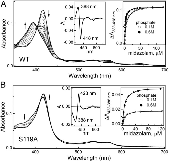
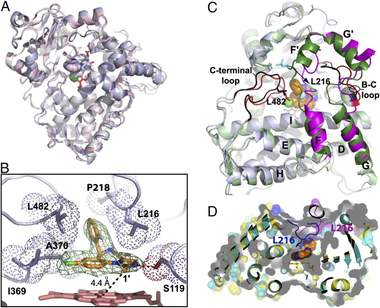
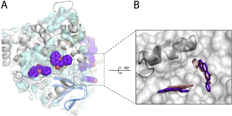
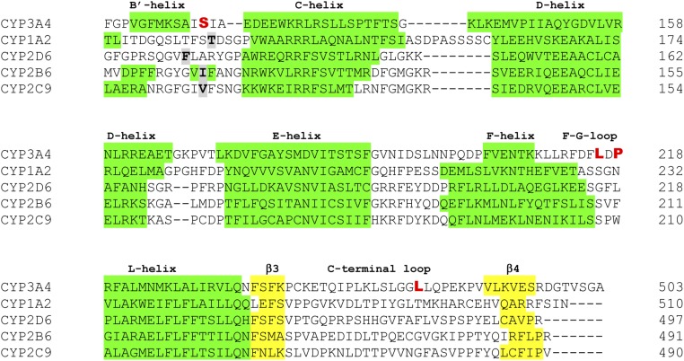
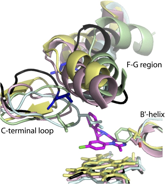
Human cytochrome P450 3A4 (CYP3A4) is a major hepatic and intestinal enzyme that oxidizes more than 60% of administered therapeutics. Knowledge of how CYP3A4 adjusts and reshapes the active site to regioselectively oxidize chemically diverse compounds is critical for better understanding structure-function relations in this important enzyme, improving the outcomes for drug metabolism predictions, and developing pharmaceuticals that have a decreased ability to undergo metabolism and cause detrimental drug-drug interactions. However, there is very limited structural information on CYP3A4-substrate interactions available to date. Despite the vast variety of drugs undergoing metabolism, only the sedative midazolam (MDZ) serves as a marker substrate for the in vivo activity assessment because it is preferentially and regioselectively oxidized by CYP3A4. We solved the 2.7 Å crystal structure of the CYP3A4-MDZ complex, where the drug is well defined and oriented suitably for hydroxylation of the C1 atom, the major site of metabolism. This binding mode requires H-bonding to Ser119 and a dramatic conformational switch in the F-G fragment, which transmits to the adjacent D, E, H, and I helices, resulting in a collapse of the active site cavity and MDZ immobilization. In addition to providing insights on the substrate-triggered active site reshaping (an induced fit), the crystal structure explains the accumulated experimental results, identifies possible effector binding sites, and suggests why MDZ is predominantly metabolized by the CYP3A enzyme subfamily.

Keywords: CYP3A4; crystal structure; drug metabolism; midazolam; substrate binding.


Conflict of interest statement

The authors declare no conflict of interest.

Figures

Fig. 1.
Fig. 1.
Structure of MDZ with the indicated primary (C1) and secondary (C4) hydroxylation sites.
Fig. 2.
Fig. 2.
(A and B) Spectral changes induced by MDZ in the wild type and S119A CYP3A4, respectively. Arrows indicate direction of absorbance changes. (Left insets) Difference spectra between the ligand-free and MDZ-bound forms. (Right insets) Titration plots and hyperbolic fittings from which the Kd values were derived. A low-to-high spin shift in CYP3A4 S119A induced by testosterone is shown in Fig. S2.
Fig. 3.
Fig. 3.
Crystal structure of the CYP3A4–MDZ complex. (A) Superposition of three molecules present in the asymmetric unit shows their similar fold (root-mean-square deviation between the ordered Cα-atoms is 0.6–0.7 Å). (B) Close-up view at the active site of molecule A. MDZ is bound above the heme plane with the C1–Fe distance of 4.4 Å and forms a hydrogen bond with Ser119 via the imidazole ring nitrogen. The chlorophenyl ring is within the Van der Waals distance from the Ile369–Ala370 fragment, whereas the fluorophenyl ring is flanked by the Leu216, Pro218, and Leu482 side chains. These interactions immobilize MDZ and orient suitably for hydroxylation of the C1-atom, the primary oxidation site. Simulated annealing Fo–Fc composite omit map for MDZ (in green mesh) is contoured at 3σ. (C) Superposition of the ligand-free (Protein Data Bank ID 1TQN; in light/dark green and black) and MDZ-bound (in gray, magenta and red) structures. Structural elements undergoing reorganization are labeled. MDZ is in orange and space-filling representation. A 12-Å shift of Leu216 (shown in cyan) is indicated by an arrow. (D) Slice through the superimposed 1TQN (cyan and magenta) and MDZ-bound structures (yellow and blue) with semitransparent surfaces to show that the substrate-induced conformational switch leads to the active site collapse.
Fig. S1.
Fig. S1.
(A) Brown color of crystalline MDZ-bound CYP3A4 indicates that the heme iron is in a high-spin form. (B) The CYP3A4–MDZ complex crystallizes in the P212121 space group, with three molecules per asymmetric unit. MDZ is shown in magenta and space-filling representation. (C) Average B-factors (for the main and side chain atoms) plotted vs. residue number of each peptide chain. Average B-factors for MDZ are listed in Table S1.
Fig. S2.
Fig. S2.
A low-to-high spin conversion in CYP3A4 S119A (0.9 μM) induced by a 40- and 80-fold excess of testosterone.
Fig. 4.
Fig. 4.
(A and B) Side view and top view, respectively, at the active site of the CYP3A4–MDZ complex, where residues mutated by Khan et al. (21) are highlighted. Residues located within the Van der Waals distance or more remotely from MDZ (in orange) are depicted in green and cyan, respectively. Some secondary structure elements were deleted to provide a better view.
Fig. 5.
Fig. 5.
(A) Reorganization in the hydrophobic cluster (in cyan) near the MDZ-flanking Leu216 (in blue) assists the 1-OH MDZ orientation. Corresponding residues from the ligand-free 1TQN structure are shown in black. (B) Location of CYP3A4 SNVs whose functional effects on the MDZ metabolism were investigated. The I and E helices are in beige and pink, respectively; the F–G fragment is in green. (C and D) Residues comprising the peripheral progesterone binding site (in different colors) and the F–G fragment (in black) in the ligand-free and MDZ-bound CYP3A4, respectively.
Fig. 6.
Fig. 6.
Possible ANF binding sites in the MDZ-bound CYP3A4. (A) Surface clefts on the proximal face with manually docked ANF molecules (in purple). The F–G fragment is in green; MDZ in orange. Potential ANF binding sites on the distal side are shown in Fig. S3. (B) Side view at the pockets, occupation of which by ANF could modulate conformational dynamics in the F–G fragment (in gray and cyan) and the Leu216-cluster formation. Selected residues comprising the potential ANF binding sites are shown in yellow sticks.
Fig. S3.
Fig. S3.
(A) Distal face of CYP3A4 with two potential α-naphthoflavone (ANF) binding sites. Cavities were displayed using the detection cutoff of 20 solvent radii, and ANF (in purple) was docked manually. It is generally accepted that the central groove on the P450 distal face serves as a redox partner docking site. By occupying this site, ANF and other effectors could modulate redox partner binding and, as a consequence, an electron flow to the heme. Two other hydrophobic pockets, occupation of which by ANF could affect MDZ metabolism, are indicated by a dashed rectangle and discussed in the main text. (B) Side view on the highlighted area near the F–G fragment in the ligand-free CYP3A4 (1TQN structure). Surface clefts preexist, but one (on the right) is deeper and the other is shallower, relative to those in the CYP3A4–MDZ complex (see Fig. 6B). ANF could bind to both sites regardless of whether the drug is bound or not, but in a different orientation. ANF molecules manually docked to the ligand-free and MDZ-bound CYP3A4 are displayed in orange and purple, respectively.
Fig. S4.
Fig. S4.
Structure-based sequence alignment of human drug-metabolizing CYPs. Only fragments containing residues critical for MDZ binding (depicted in bold red) are shown. The B′–C peptide residues occupying the CYP3A4 Ser119 position (shown in Fig. S5) are highlighted in black/gray.
Fig. S5.
Fig. S5.
Structural overlay of human drug-metabolizing CYPs: CYP3A4 (1WOE; black), CYP1A2 (2HI4; cyan), CYP2B6 (molecule A of 4RQL; pink), CYP2C9 (molecule A of 1OG2; yellow), and CYP2D6 (molecule A of 4WNU; green). MDZ (in magenta) is shown in the crystallographic binding mode. Neither of the B′–C loop residues aligning with Ser119 of CYP3A4 can H-bond and accommodate MDZ in a similar mode. The length and topology of the F–G and C-terminal loops significantly varies among CYPs, due to which it is not possible to accurately pinpoint residues corresponding to MDZ-flanking leucines 216 and 482. Nevertheless, the outlined sequence and structural dissimilarities, and preservation of all key elements in CYP3A5 and CYP3A7 (Fig. S6) could explain why MDZ is predominantly oxidized by the CYP3A enzyme subfamily.
Fig. S6.
Fig. S6.
Amino acid sequence alignment of the CYP3A enzyme subfamily preformed with Clustal Omega. Residues critical for MDZ binding are highlighted in bold red.

References

    1. Guengerich FP. Cytochrome P-450 3A4: Regulation and role in drug metabolism. Annu Rev Pharmacol Toxicol. 1999;39:1–17. - PubMed
    1. Rendic S, Di Carlo FJ. Human cytochrome P450 enzymes: A status report summarizing their reactions, substrates, inducers, and inhibitors. Drug Metab Rev. 1997;29(1-2):413–580. - PubMed
    1. Atkins WM. Non-Michaelis-Menten kinetics in cytochrome P450-catalyzed reactions. Annu Rev Pharmacol Toxicol. 2005;45:291–310. - PubMed
    1. Yano JK, et al. The structure of human microsomal cytochrome P450 3A4 determined by X-ray crystallography to 2.05-A resolution. J Biol Chem. 2004;279(37):38091–38094. - PubMed
    1. Williams PA, et al. Crystal structures of human cytochrome P450 3A4 bound to metyrapone and progesterone. Science. 2004;305(5684):683–686. - PubMed

Publication types

MeSH terms

Associated data