. 2017 Jan 17;114(3):562-567.
doi: 10.1073/pnas.1612195114. Epub 2016 Dec 28.

CD74 is a novel transcription regulator

Affiliations

CD74 is a novel transcription regulator

Naama Gil-Yarom et al. Proc Natl Acad Sci U S A. .

Abstract

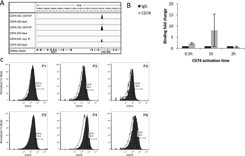
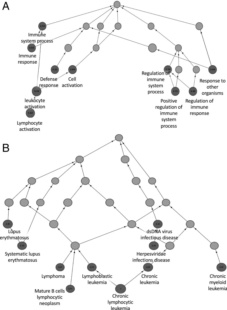
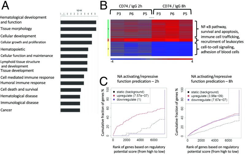
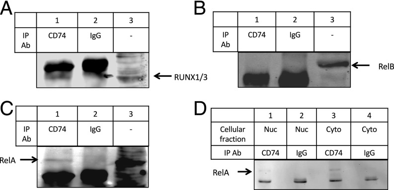
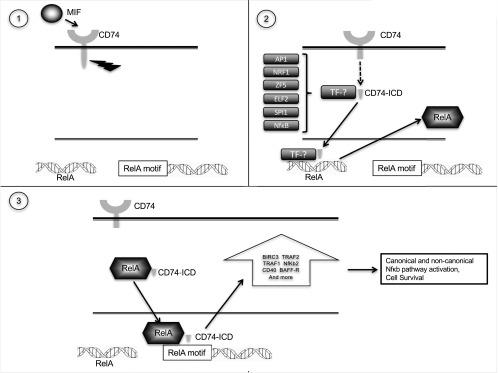
CD74 is a cell-surface receptor for the cytokine macrophage migration inhibitory factor. Macrophage migration inhibitory factor binding to CD74 induces its intramembrane cleavage and the release of its cytosolic intracellular domain (CD74-ICD), which regulates cell survival. In the present study, we characterized the transcriptional activity of CD74-ICD in chronic lymphocytic B cells. We show that following CD74 activation, CD74-ICD interacts with the transcription factors RUNX (Runt related transcription factor) and NF-κB and binds to proximal and distal regulatory sites enriched for genes involved in apoptosis, immune response, and cell migration. This process leads to regulation of expression of these genes. Our results suggest that identifying targets of CD74 will help in understanding of essential pathways regulating B-cell survival in health and disease.

Keywords: CD74; CLL; NF-κB; RUNX; transcription.


Conflict of interest statement

The authors declare no conflict of interest.

Figures

Fig. S1.
Fig. S1.
CD74 binding to the chromatin. (A) 293T cells were transfected with CD74–ICD–myc plasmid. ChIP-seq was done using CD74 or myc antibodies. Binding of CD74 in the proximity of the NLE1 gene was detected with both antibodies. (B) CLL cells were activated with anti-CD74 Ab for 0.5 h, 1 and 2 h. Following activation, the cells were fixed, and Chip-RT was conducted using CD74 Ab. qRT-PCR of a binding area in the proximity of the NLE1 gene was used to determine binding levels of CD74 to the chromatin in CLL cells. Binding of CD74–ICD to the chromatin is detected 0.5 and 1 h following activation (n = 2). (C) CD74 cell-surface expression on CLL cells was determined by FACS analysis.
Fig. 1.
Fig. 1.
CD74–ICD binds the chromatin in regulatory areas. CLL cells from four different samples were stimulated for 1 h with anti-CD74 Ab. ChIP-seq was then performed using anti-CD74 or isotype control antibodies. Binding of CD74 to the chromatin was compared with the input. (A) A representative analysis of binding of CD74–ICD to a promoter region. ChIP-Seq peaks are shown for anti-CD74 and input DNA of bigWig files of two representative experiments. (B) Binding was analyzed in all four samples and overlaps were examined. Peaks that were found in two or more samples were considered as high-confidence peaks, and their distances (kilobases) from the nearest TSS were analyzed using GREAT. (C) The distribution of important genomic features in the whole genome (Left) and in CD74 ChIP-seq peaks (Right) was obtained from CEAS. A representative analysis is shown. (D) CLL cells from two different patients were stimulated for 1 h with anti-CD74 Ab. ChIP-seq was performed with anti-H3K27Ac, anti-H3K4Me3, or anti-H3K4Me2. The diagram shows the overlap between CD74 peaks and the acetylation and methylation peaks detected by these antibodies. (E) CLL cells from two different patients were stimulated for 1 h with either anti-CD74 Ab or IgG control Ab. ChIP-seq was done with anti-H3K4Me2. The ngs.plot tool was used to plot average reads per million mapped reads (RPKM) over high-confidence CD74 peaks. The center on the x axis relates to the summits of the peaks. The SE is shaded. The diagram shows the correlation between CD74 peaks and H3K4Me2, with (orange) and without (green) CD74 activation. WCE, whole cell extract.
Fig. 2.
Fig. 2.
CD74 has a specific immune oriented role as a transcription factor. CLL cells from four samples were activated with anti-CD74 activating Ab for 1 h. Following activation, the cells were fixed, and ChIP-seq was conducted using CD74 Ab. GREAT analysis of the high confidence peaks was performed. The local directed acyclic graph is shown. The numbers relates to the binomial fold-change. (A) The biological processes controlled by CD74 identified by Gene Ontology (GO) analysis. (B) Enriched terms in disease ontology showing that CD74 specifically binds in the proximity of genes that are involved in diseases relating to lymphocytes, specifically in different types of lymphomas. dsDNA, double-stranded DNA.
Fig. 3.
Fig. 3.
CD74 activation leads to transcription regulation. Samples from three patients were activated with either anti-CD74 or control IgG antibodies for 2 and 8 h. mRNA was extracted and subjected to mRNA sequencing. mRNA levels were compared between CD74- and IgG-activated cells. (A) Differentially expressed genes at the 8-h time point (Padj < 0.05, absolute fold-change > 2, reads count of at least 20 in at least one sample) were analyzed using the Ingenuity system. Log2 ratios of CD74 activation versus IgG are depicted in a color scale. (B) Differentially expressed genes in response to CD74 activation after 2 or 8 h (total of 603 genes) were clustered to three clusters using Euclidean distance measure. For each patient and time point, log2 ratios of CD74 activation versus IgG is shown. Cluster 1 includes genes that are up-regulated at 2 h, and more strongly up-regulated at 8 h; cluster 2 is characterized by genes that were up-regulated shortly after activation, and remained at high levels at the later time point; and cluster 3, in which the genes are down-regulated at 2 h, with stronger down-regulation seen at 8 h. Enriched pathways are indicated near each cluster. (C) BETA software was used to correlate between the genes that were bound in their proximity by CD74, and genes that were regulated at the mRNA level.
Fig. 4.
Fig. 4.
CD74 forms a complex with other transcription factors to regulate transcription. CLL cells were activated for 1 h with CD74 Ab. Total proteins were extracted, and immunoprecipitation was performed with anti-CD74 or isotype control antibodies. Proteins were separated on a 10% gel, and immunoblots were analyzed for RUNX1 and RUNX3 (A), RelB (B), and RelA (C). (D) CLL cells were activated for 1 h with anti-CD74 Ab. Nuclear and cytoplasmic fractions were separated using the Nucbuster extraction kit. Immunoprecipitation was performed using CD74 Ab (lanes 1 and 3) or IgG control Ab (lanes 2 and 4). Immunoprecipitates were separated on 10% SDS/PAGE, and analyzed for RelA.
Fig. 5.
Fig. 5.
CD74 up-regulates expression of genes that are part of the NF-κB–induced survival cascade. (A and B) CLL cells from three patients were activated with anti-CD74 Ab for 2 and 8 h. (A) mRNA-seq showing elevation in genes that take part in the NF-κB survival pathway. For B-cell activating factor (BAFF)-R level determination, CLL cells were activated with CD74 Ab for 18 h. mRNA was extracted, and BAFF-R levels were determined using qRT-PCR (n = 4, P < 0.05). (B) mRNA-seq showing elevation in genes that are Runx1/3 targets. (C) TRAF1 and BIRC3 protein levels 48 h following activation were analyzed by FACS. Graph presents fold-change relative to unactivated cells (n = 4, P < 0.01; n = 6, P < 0.05). (D) CLL cells (5 × 10⁶) were transfected with siRNA against CD74 or control siRNA. After 24 h, cells were harvested, RNA was purified and mRNA levels of TRAF and BIRC3 were analyzed by qRT-PCR (n = 7, *P = 0.047 for TRAF, P = 0.0258 for BIRC3). *P ≤ 0.05; **P ≤ 0.01; ***P ≤ 0.001; ****P ≤ 0.0001.
Fig. S2.
Fig. S2.
A summarizing model. TF, transcription factor.

References

    1. Stumptner-Cuvelette P, Benaroch P. Multiple roles of the invariant chain in MHC class II function. Biochim Biophys Acta. 2002;1542(1-3):1–13. - PubMed
    1. Arneson LS, Miller J. The chondroitin sulfate form of invariant chain trimerizes with conventional invariant chain and these complexes are rapidly transported from the trans-Golgi network to the cell surface. Biochem J. 2007;406(1):97–103. - PMC - PubMed
    1. Bucala R, Shachar I. The integral role of CD74 in antigen presentation, MIF signal transduction, and B cell survival and homeostasis. Mini Rev Med Chem. 2014;14(14):1132–1138. - PubMed
    1. Cohen S, Shachar I. Cytokines as regulators of proliferation and survival of healthy and malignant peripheral B cells. Cytokine. 2012;60(1):13–22. - PubMed
    1. Matza D, Kerem A, Medvedovsky H, Lantner F, Shachar I. Invariant chain-induced B cell differentiation requires intramembrane proteolytic release of the cytosolic domain. Immunity. 2002;17(5):549–560. - PubMed

Publication types