

Clinical and histological evaluation of large macular hole surgery using the inverted internal limiting membrane flap technique
- PMID: 28031697
- PMCID: PMC5179209
- DOI: 10.2147/OPTH.S119762
Clinical and histological evaluation of large macular hole surgery using the inverted internal limiting membrane flap technique
Abstract
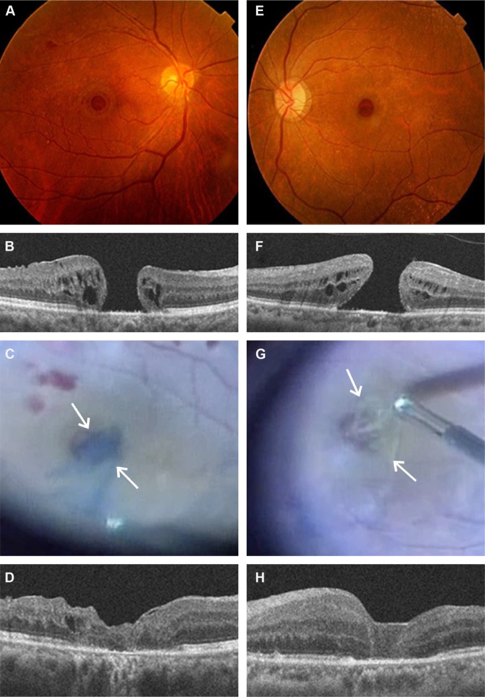
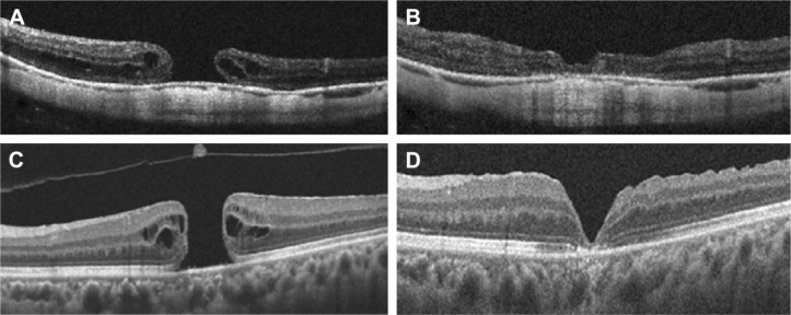
Purpose: The aims of this study were to analyze optical coherence tomography (OCT) imaging of large macular holes (MHs) treated with inverted internal limiting membrane (ILM) flap technique and to perform a histological examination of an ILM-like membrane tissue obtained during vitrectomy.
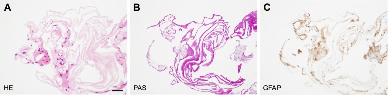
Patients and methods: This is a retrospective observational case study. Nine patients, comprising of five males and four females, showing large and myopic MHs, underwent pars plana vitrectomy (PPV) with inverted ILM flap technique assisted by brilliant blue G (BBG) staining. Ophthalmological findings including visual acuity and OCT were investigated based on medical records. Formalin-fixed paraffin-embedded tissue section of an ILM-like membrane was submitted for immunohistochemistry with glial fibrillary acidic protein (GFAP).
Results: ILM was clearly stained with BBG in eight patients, whereas the ILM in one case revealed no staining with BBG during PPV. Visual acuities improved to >0.2 LogMAR in six patients. The complete closure of MH following PPV with inverted ILM technique was eventually achieved in all patients determined by OCT imaging (100%). Only one patient showed recovery of ellipsoid zone and interdigitation zone following the surgery. Elongation of outer nuclear layer was noted in three eyes. The ILM-like membrane not stained with BBG histologically revealed an amorphous structure admixed with GFAP-positive mononuclear cell infiltration.
Conclusion: PPV with inverted ILM flap technique achieved 100% closure rates with favorable configuration at an initial surgery in large MHs. Our histopathological data also suggest that even BBG staining-negative membrane may be a useful material for autologous transplantation to the hole.
Keywords: OCT; glial cells; histopathology; inverted ILM flap; macular hole.
Conflict of interest statement
The authors report no conflicts of interest in this work.
Figures
References
-
- Kase S, Saito W, Ohno S, Ishida S. Cyclo-oxygenase-2 expression in human idiopathic epiretinal membrane. Retina. 2010;30(5):719–723. - PubMed
-
- Schumann RG, Eibl KH, Zhao F, et al. Immunocytochemical and ultrastructural evidence of glial cells and hyalocytes in internal limiting membrane specimens of idiopathic macular holes. Invest Ophthalmol Vis Sci. 2011;52(11):7822–7834. - PubMed
-
- Steel DH, Dinah C, Madi HA, White K, Rees J. The staining pattern of brilliant blue G during macular hole surgery: a clinicopathologic study. Invest Ophthalmol Vis Sci. 2014;55(9):5924–5931. - PubMed
-
- Kuriyama S, Hayashi H, Jingami Y, Kuramoto N, Akita J, Matsumoto M. Efficacy of inverted internal limiting membrane flap technique for the treatment of macular hole in high myopia. Am J Ophthalmol. 2013;156:125–131.e1. - PubMed
LinkOut - more resources
Full Text Sources
Other Literature Sources
Medical
Miscellaneous
