. 2016 Dec 20:4:94.
doi: 10.1186/s40425-016-0199-9. eCollection 2016.

Sarcoidosis in the setting of combination ipilimumab and nivolumab immunotherapy: a case report & review of the literature

Affiliations

Sarcoidosis in the setting of combination ipilimumab and nivolumab immunotherapy: a case report & review of the literature

Joshua E Reuss et al. J Immunother Cancer. .

Abstract

Background: We report a case of sarcoidosis in a patient with metastatic melanoma managed with combination ipilimumab/nivolumab. Sarcoid development has been linked with single agent immunotherapy but, to our knowledge, it has not been reported with combination ipilimumab/nivolumab treatment. This case raises unique management challenges for both the melanoma and the immunotherapy-related toxicity.

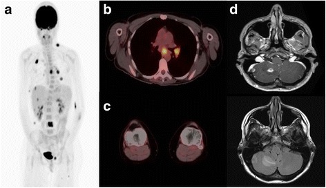
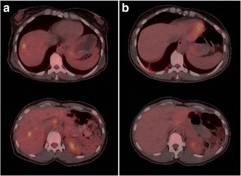
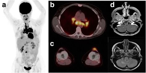
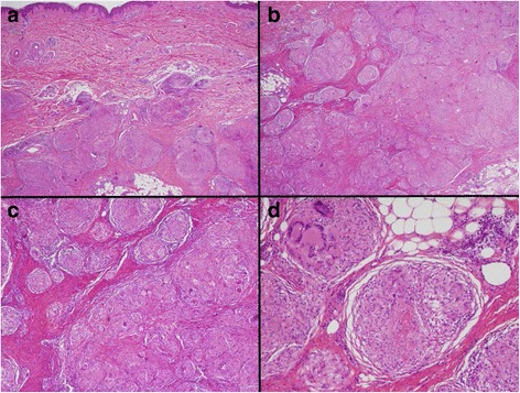
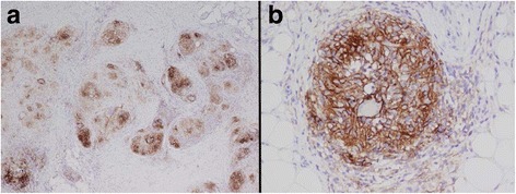
Case presentation: A 46 year old Caucasian female with M1c-metastatic melanoma was managed with ipilimumab/nivolumab combination. Patient experienced response in baseline lesions but developed new clinical and radiographic findings. Biopsy of new lesions at two different sites both demonstrated tumefactive sarcoidosis. Staining of the biopsy tissue for PD-L1 expression demonstrated strong PD-L1 staining of the histiocytes and lymphocytes within the granulomas. Monotherapy nivolumab was continued without progression of sarcoid findings or clinical deterioration.

Conclusions: Tissue biopsy for evaluation of new lesions on immunotherapy is an important step to help guide decision making, as non-melanoma lesions can mimic disease progression.

Keywords: Checkpoint inhibitor; Combination immunotherapy; Immune-related adverse event; Ipilimumab; Metastatic melanoma; Nivolumab; Sarcoidosis.


Figures

Fig. 1
Fig. 1
Imaging at Time of Diagnosis Before Treatment. a Scout PET scan showing cervical/mediastinal/inguinal adenopathy and vertebral lesions. b Cross section of mediastinum with increased update in mediastinal nodes (SUVmax 10 in subcarinal node). c Cross section of knees with no increased uptake. d MRI brain with two right cerebellar metastases with vasogenic edema
Fig. 2
Fig. 2
PET-CT of Hepatic Metastases Before and After Combination Immunotherapy. a PET-CT scan at times of diagnosis of with several hypermetabolic hepatic lesions consistent with metastases. b PET-CT scan after 4 cycles of nivolumab plus ipilimumab and 3 cycles of nivolumab maintenance
Fig. 3
Fig. 3
PET-CT scan after Nivolumab + Ipilimumab Induction and on Maintenance Nivolumab. a Scout PET scan showing cervical/mediastinal/inguinal adenopathy and vertebral lesions. b Cross section of mediastinum with increased uptake in mediastinal nodes (SUVmax 16 in paratracheal node). c Cross section of knees with increased uptake in left soft tissue nodule (SUVmax 2.7). d MRI brain with interval improvement of right cerebellar metastases
Fig. 4
Fig. 4
Hematoxylin and Eosin Staining of Pre-Tibial Soft Tissue Nodule. Microscopic hematoxylin and eosin (H&E) section showing (A) large, deep dermal collections of non-caseating granulomas (a, 2×) extending into the subcutaneous tissue (b, 2×). Higher magnification shows tightly formed granulomas containing multinucleated giant cells separated by fibrous connective tissue (c, 4×). Scattered mature appearing lymphocytes are seen surrounding the granulomatous inflammation (d, 10×)
Fig. 5
Fig. 5
PD-L1 Staining of Pre-Tibial Soft Tissue Nodule. PD-L1 antibody stained section of the granulomatous inflammation shows strong membranous staining of the histiocytes within the granulomas and scattered positive lymphocytes (a, 4×; b, 20×)

References

    1. Hodi FS, O’Day SJ, McDermott DF, Weber RW, et al. Improved survival with ipilimumab in patients with metastatic melanoma. N Engl J Med. 2010;363(8):711–23. doi: 10.1056/NEJMoa1003466. - DOI - PMC - PubMed
    1. Postow MA, Chesney J, Pavlick AC, Robert C, et al. Nivolumab and ipilimumab versus ipilimumab in untreated melanoma. N Engl J Med. 2015;372(21):2006–17. doi: 10.1056/NEJMoa1414428. - DOI - PMC - PubMed
    1. Larkin J, Chiarion-Sileni V, Gonzalez R, Grob JJ, et al. Combined nivolumab and ipilimumab or monotherapy in untreated melanoma. N Engl J Med. 2015;373(1):23–34. doi: 10.1056/NEJMoa1504030. - DOI - PMC - PubMed
    1. Robert C, Schachter J, Long GV, Arance A, et al. Pembrolizumab versus Ipilimumab in Advanced Melanoma. N Engl J Med. 2015;372(26):2521–32. doi: 10.1056/NEJMoa1503093. - DOI - PubMed
    1. Eckert A, Schoeffler A, Dalle S, Phan A, et al. Anti-CTLA4 monoclonal antibody induced sarcoidosis in a metastatic melanoma patient. Dermatology. 2009;218(1):69–70. doi: 10.1159/000161122. - DOI - PubMed

LinkOut - more resources