

Nanomedicine as a strategy to fight thrombotic diseases
- PMID: 28031907
- PMCID: PMC5137979
- DOI: 10.4155/fso.15.46
Nanomedicine as a strategy to fight thrombotic diseases
Abstract
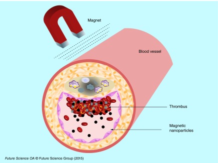
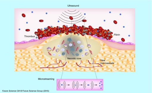
This review highlights the preclinical and clinical research based on the use of nano- and micro-carriers in thrombolytic drug delivery. Ischemic heart and stroke caused by thrombosis are the main causes of death in the world. Because of their inactivation in the blood, high doses of thrombolytics are administered to patients, increasing the risk of intracranial hemorrhage. Preclinical research conducted with lipid, polymer or magnetic nanoparticles loaded with thrombolytic drugs showed an enhancement of thrombolysis and a reduction of undesirable side effects. Targeted nanocarriers exhibited an increased accumulation into clot. Clinical trials were already conducted with lipid-based microbubbles combined with ultrasound and thrombolytic drug and showed thrombolysis improvement. Future validation of nanosystems is awaited in clinic. This research opens new strategies for the management of thrombotic diseases.
Keywords: animal models; drug delivery; ischemic heart; microbubbles; nanocarriers; stroke; thrombolytic.
Conflict of interest statement
Financial & competing interests disclosure This work was supported by Lefoulon-Delalande scholarships (to M Varna and R Bayles), EU project NanoAthero FP7-NMP-2012-LARGE-6–309820, IMOVA project (FUI/OSEO, CG93), and ANR-13-LAB1–0005–01 ‘FucoChem’, University Paris 13 and Inserm. The authors have no other relevant affiliations or financial involvement with any organization or entity with a financial interest in or financial conflict with the subject matter or materials discussed in the manuscript apart from those disclosed. No writing assistance was utilized in the production of this manuscript.
Figures
References
-
- Hilgendorf I, Swirski FK, Robbins CS. Monocyte fate in atherosclerosis. Arterioscler. Thromb. Vasc. Biol. 2015;35(2):272–279. - PubMed
-
- Falk E. Pathogenesis of atherosclerosis. J. Am. Coll. Cardiol. 2006;47(8 Suppl.):C7–C12. - PubMed
-
- Fuster V, Fayad ZA, Moreno PR, Poon M, Corti R, Badimon JJ. Atherothrombosis and high-risk plaque: part ii: approaches by noninvasive computed tomographic/magnetic resonance imaging. J. Am. Coll. Cardiol. 2005;46(7):1209–1218. - PubMed
Publication types
LinkOut - more resources
Full Text Sources
Other Literature Sources