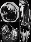
Pigmented villonodular synovitis of proximal tibiofibular joint: Rare site of involvement treated with medical management
- PMID: 28032093
- PMCID: PMC5184763
- DOI: 10.4103/2278-330X.195348
Pigmented villonodular synovitis of proximal tibiofibular joint: Rare site of involvement treated with medical management
Conflict of interest statement
There are no conflicts of interest.
Figures


Similar articles
-
Localized pigmented villonodular synovitis of the proximal tibiofibular joint.Knee Surg Relat Res. 2014 Dec;26(4):249-52. doi: 10.5792/ksrr.2014.26.4.249. Epub 2014 Dec 2. Knee Surg Relat Res. 2014. PMID: 25505708 Free PMC article.
-
Pigmented villonodular synovitis of the proximal tibiofibular joint.Australas Radiol. 2004 Dec;48(4):520-2. doi: 10.1111/j.1440-1673.2004.01371.x. Australas Radiol. 2004. PMID: 15601334
-
Pigmented villonodular synovitis of the distal tibiofibular joint: a case report.Clin Podiatr Med Surg. 2011 Jun;28(3):589-97. doi: 10.1016/j.cpm.2011.04.005. Epub 2011 Jun 8. Clin Podiatr Med Surg. 2011. PMID: 21777788
-
Pigmented villonodular synovitis of the temporomandibular joint.Arch Otolaryngol Head Neck Surg. 1997 May;123(5):536-9. doi: 10.1001/archotol.1997.01900050092012. Arch Otolaryngol Head Neck Surg. 1997. PMID: 9158403 Review.
-
[Unusual presentation of pigmented villonodular synovitis of the hip joint: case report and review of the literature].Acta Ortop Mex. 2017 Nov-Dec;31(6):308-311. Acta Ortop Mex. 2017. PMID: 29641859 Review. Spanish.
Cited by
-
Pigmented Villonodular Synovitis of Thumb-A Cytological Diagnosis.J Clin Diagn Res. 2017 Jun;11(6):ED18-ED20. doi: 10.7860/JCDR/2017/28184.10099. Epub 2017 Jun 1. J Clin Diagn Res. 2017. PMID: 28764182 Free PMC article.
References
-
- Murphey MD, Rhee JH, Lewis RB, Fanburg-Smith JC, Flemming DJ, Walker EA. Pigmented villonodular synovitis: Radiologic-pathologic correlation. Radiographics. 2008;28:1493–518. - PubMed
-
- Garner HW, Ortiguera CJ, Nakhleh RE. Pigmented villonodular synovitis. Radiographics. 2008;28:1519–23. - PubMed
-
- Sheldon PJ, Forrester DM, Learch TJ. Imaging of intraarticular masses. Radiographics. 2005;25:105–19. - PubMed
-
- Hughes TH, Sartoris DJ, Schweitzer ME, Resnick DL. Pigmented villonodular synovitis: MRI characteristics. Skeletal Radiol. 1995;24:7–12. - PubMed
-
- Ryan RS, Louis L, O’Connell JX, Munk PL. Pigmented villonodular synovitis of the proximal tibiofibular joint. Australas Radiol. 2004;48:520–2. - PubMed
LinkOut - more resources
Full Text Sources
Other Literature Sources

