

Relationship between Patellar Tracking and the "Screw-home" Mechanism of Tibiofemoral Joint
- PMID: 28032709
- PMCID: PMC6584384
- DOI: 10.1111/os.12295
Relationship between Patellar Tracking and the "Screw-home" Mechanism of Tibiofemoral Joint
Abstract
Objective: To demonstrate the effect of the screw-home motion on the stability of the patellofemoral joint, and investigate its mechanism of regulation of patellar tracking.
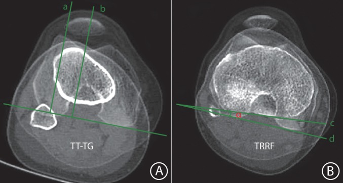
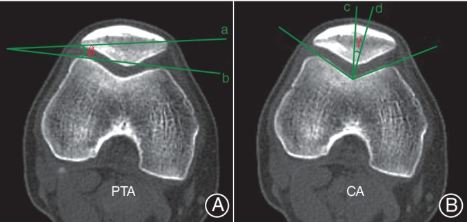
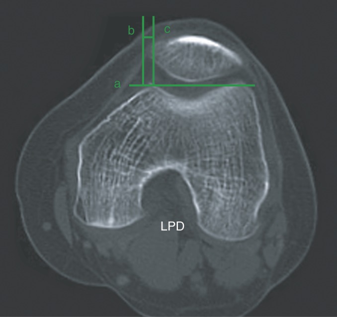
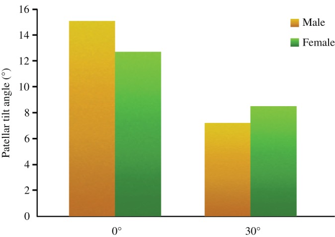
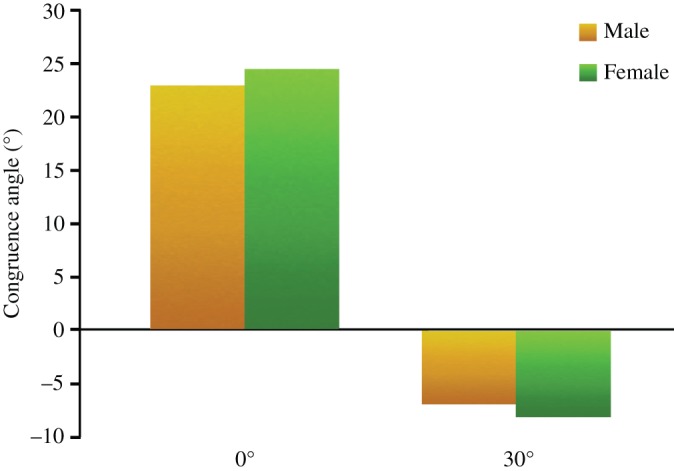
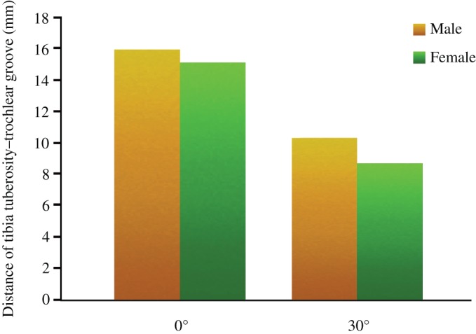
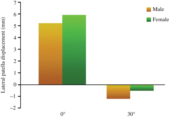
Methods: Twenty volunteers who met the criteria were examined. All subjects had axial computed tomography (CT) scanning performed on bilateral knees at 0° and 30° of flexion. Scanning began above the femorotibial articulation and femoral trochlear groove, and moved sequentially down to the level of the anterior tibial tubercle. The following measurements were obtained: tibial rotation relative to the femur (TRRF), tibial tuberosity-trochlear groove (TT-TG) distance, lateral patellar displacement (LPD), patellar tilt angle (PTA), and congruence angle (CA). We assessed the change (Δ) in each variable at both flexion angles, and analyzed this to investigate the corresponding relationship between the patella, the femur, and the screw-home mechanism. The differences between the values measured at 0° and those measured at 30° flexion were analyzed using the paired sample t-test. The differences between men and women were analyzed using the t-test. Pearson's correlations were performed to determine the relationship between ΔTT-TG distance and ΔLPD, ΔPTA and ΔTRRF, and ΔCA and ΔTRRF.
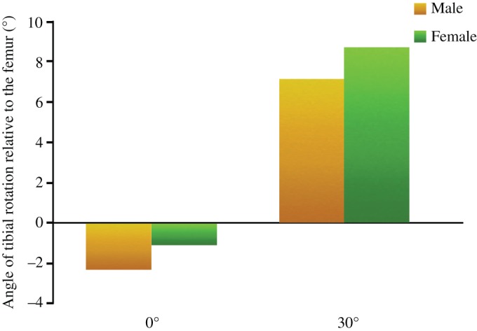
Results: There were 10 women and 10 men enrolled in the present study, with an average age of 25 years and an average body mass index of 21.8 kg/m2 , and all volunteers had no history of knee injuries. Compared with measurements taken at 0° flexion, TRRF at 30° flexion was significantly increased, and the PTA, CA, LPD, and TT-TG distance were significantly decreased (all P < 0.01). There was no difference between men and women at 0° and 30° flexion, respectively (P < 0.01). In this respect, there was no sex difference, but the change was greater for men than for women. Both ΔPTA and ΔCA demonstrated significant correlation with the ΔTRRF (both P < 0.01); a significant correlation between ΔLPD and ΔTT-TG distance was also demonstrated (P < 0.01).
Conclusions: As the tibiofemoral joint rotated, the patellofemoral joint became more stable and aligned, which indicates that the screw-home mechanism plays an important role in regulating patellofemoral joint alignment.
Keywords: Kinematics; Patellar tracking; Patellofemoral joint; Tibiofemoral joint; “Screw-home” mechanism.
© 2016 Chinese Orthopaedic Association and John Wiley & Sons Australia, Ltd.
Figures
References
-
- Distefano MD, Michael C. Disorders of the patellofemoral joint. 4th edn. J Bone Joint Surg Br, 2005, 87: 482.
-
- Senavongse W, Amis AA. The effects of articular, retinacular, or muscular deficiencies on patellofemoral joint stability: a biomechanical study in vitro. J Bone Joint Surg Br, 2005, 87: 577–582. - PubMed
-
- Katchburian MV, Bull AM, Shih YF, Heatley FW, Amis AA. Measurement of patellar tracking: assessment and analysis of the literature. Clin Orthop Relat Res, 2003, 412: 241–259. - PubMed
MeSH terms
LinkOut - more resources
Full Text Sources
Other Literature Sources
Research Materials
Miscellaneous
