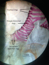
Calcification of the lower respiratory tract in relation to flight development in Jamaican fruit bats (Phyllostomidae, Artibeus jamaicensis)
- PMID: 28033680
- PMCID: PMC5345678
- DOI: 10.1111/joa.12575
Calcification of the lower respiratory tract in relation to flight development in Jamaican fruit bats (Phyllostomidae, Artibeus jamaicensis)
Abstract
The production of echolocation calls in bats along with forces produced by contraction of thoracic musculature used in flight presumably puts relatively high mechanical loads on the lower respiratory tract (LRT). Thus, there are likely adaptations to prevent collapse or distortion of the bronchial tree and trachea during flight in echolocating bats. By clearing and staining (Alcian blue and Alizarin red) LRTs removed from nonvolant neonates, semivolant juveniles, volant subadults, and adult Jamaican fruit bats (Artibeus jamaicensis), I found that calcification of the tracheal, primary bronchial, and secondary bronchial (lobar) cartilage rings occurs over the span of about 3 days and coincides with later developmental stages of flight and the increased production of echolocation calls. Tracheal rings that are immediately adjacent to the larynx calcified first, followed by more caudal tracheal rings and then the rings of the primary and secondary bronchi. I suggest that calcification of LRT cartilage rings in echolocating bats provides increased rigidity to counter the thoracic compressions incurred during flight. Calcification of the LRT rings is an adaptation to support the emission of laryngeally produced echolocation calls during flight in bats.
Keywords: bats; bronchi; calcification; trachea.
© 2016 Anatomical Society.
Figures


References
-
- Adams RA (2009) Methods for assessing prenatal growth and development in bats In: Ecological and Behavioral Methods for the Study of Bats, 2nd edn (eds. Kunz TH, Parsons S.), pp. 265–272. Baltimore: The Johns Hopkins University Press.
-
- Adams RA, Shaw JB (2013) Time's arrow and the evolutionary development of bat flight In: Bat Evolution, Ecology and Conservation. (eds Adams RA, Pedersen SC.), pp. 21–46. New York: Springer Press.
-
- Carter RT, Adams RA (2014) Ontogeny of the larynx and flight ability in Jamaican fruit bats (Phyllostomidae) with considerations for the evolution of echolocation. Anat Rec 297, 1270–1277. - PubMed
-
- Carter RT, Shaw JB, Adams RA (2014) Ontogeny of vocalization in Jamaican fruit bats with implications for the evolution of echolocation. J Zool (Lond) 293, 25–32.
-
- Denny SP (1976) Comparative anatomy of the larynx In: The Scientific Basis of Otolaryngology. (eds Hincliffe R, Harrison DFN.), pp. 536–545. London: Heinemann.
MeSH terms
LinkOut - more resources
Full Text Sources
Other Literature Sources

