

Failure of physiologic transformation of spiral arteries, endothelial and trophoblast cell activation, and acute atherosis in the basal plate of the placenta
- PMID: 28034657
- PMCID: PMC5881902
- DOI: 10.1016/j.ajog.2016.12.029
Failure of physiologic transformation of spiral arteries, endothelial and trophoblast cell activation, and acute atherosis in the basal plate of the placenta
Abstract
Background: Failure of physiologic transformation of spiral arteries has been reported in preeclampsia, fetal growth restriction, fetal death, and spontaneous preterm labor with intact or ruptured membranes. Spiral arteries with failure of physiologic transformation are prone to develop atherosclerotic-like lesions of atherosis. There are striking parallels between preeclampsia and atherosclerotic disease, and between lesions of atherosis and atherosclerosis. Endothelial activation, identified by intercellular adhesion molecule-1 expression, is present in atherosclerotic-like lesions of heart transplantation, and is considered a manifestation of rejection. Similarly, endothelial activation/dysfunction has been implicated in the pathophysiology of atherosclerosis and preeclampsia. Intercellular adhesion molecule-1-overexpressing-activated endothelial cells are more resistant to trophoblast displacement than nonactivated endothelium, and may contribute to shallow spiral artery trophoblastic invasion in obstetrical syndromes having failure of physiologic transformation.
Objective: We sought to determine whether failure of spiral artery physiologic transformation was associated with activation of interstitial extravillous trophoblasts and/or spiral artery endothelium and presence of acute atherosis in the placental basal plate.
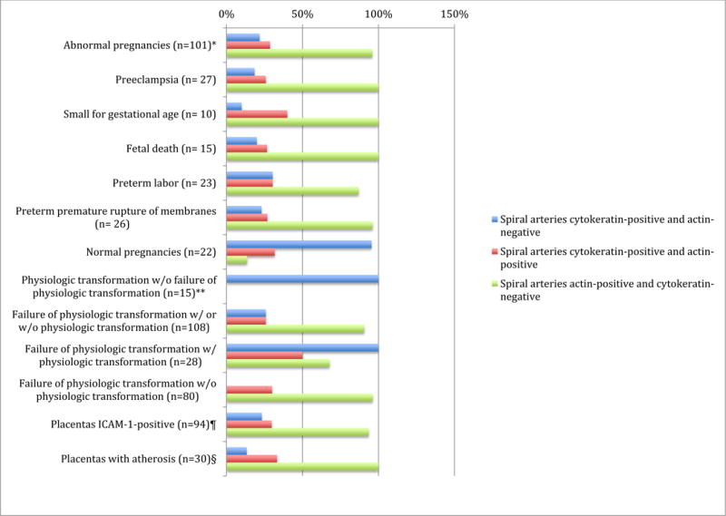
Study design: A cross-sectional study of 123 placentas (19-42 weeks' gestation) obtained from normal pregnancies (n = 22), preterm prelabor rupture of membranes (n = 26), preterm labor (n = 23), preeclampsia (n = 27), intrauterine fetal death (n = 15), and small for gestational age (n = 10) was performed. Failure of spiral artery physiologic transformation and presence of cell activation was determined using immunohistochemistry of placental basal plates containing a median of 4 (minimum: 1; maximum: 9) vessels per placenta. Endothelial/trophoblast cell activation was defined by the expression of intercellular adhesion molecule-1. Investigators examining microscopic sections were blinded to clinical diagnosis. Pairwise comparisons among placenta groups were performed with Fisher exact test and Wilcoxon rank sum test using a Bonferroni-adjusted level of significance (.025).
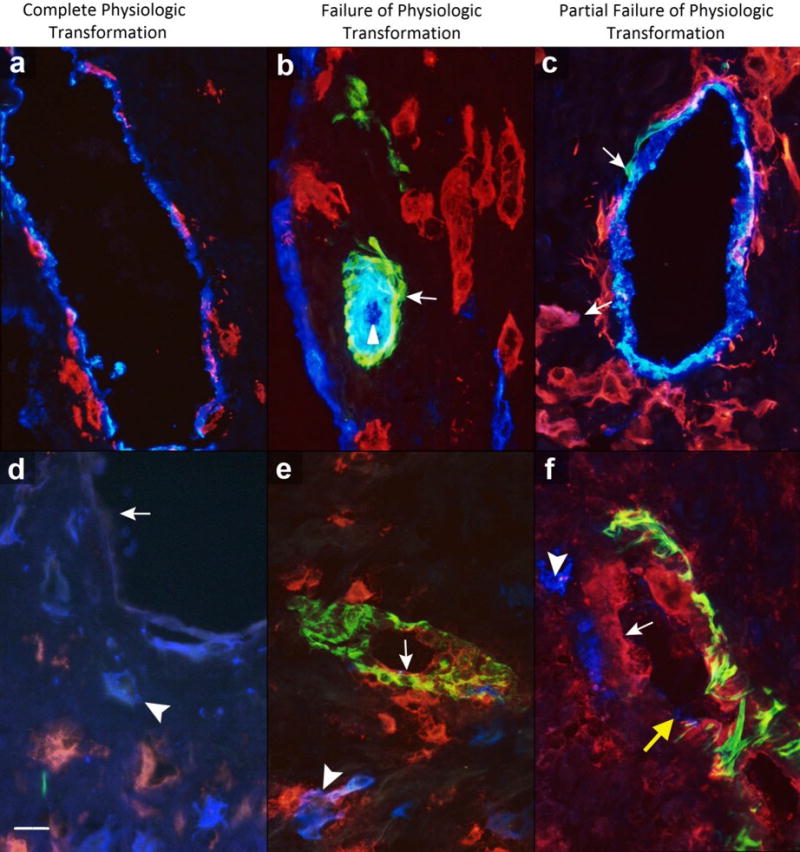
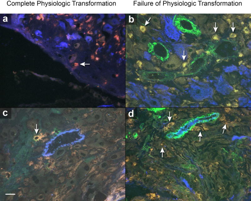
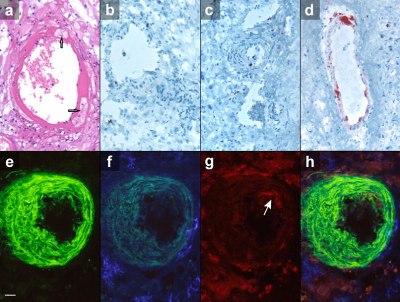
Results: We found that 87% (94/108) of placentas having spiral arteries with failure of physiologic transformation (actin-positive and cytokeratin-negative) in the basal plate, and 0% (0/15) of placentas having only spiral arteries with complete physiologic transformation (cytokeratin-positive and actin-negative), had arterial endothelial and/or interstitial extravillous trophoblasts reactive with the intercellular adhesion molecule-1 activation marker (P < .001). A significant correlation (R2 = 0.84) was found between expression of spiral artery endothelial and interstitial extravillous trophoblast intercellular adhesion molecule-1 (P < .001) in activated placentas. Lesions of atherosis were found in 31.9% (30/94) of placentas with complete and/or partial failure of physiologic transformation of spiral arteries that were intercellular adhesion molecule-1-positive, in none of the 14 placentas with failure of physiologic transformation that were intercellular adhesion molecule-1-negative, and in none of the 15 placentas with complete spiral artery physiologic transformation without failure (P = .001). All placentas (30/30, 100%) with atherosis were identified in placentas having concomitant spiral artery endothelial and interstitial extravillous trophoblast activation.
Conclusion: Failure of spiral artery physiologic transformation in the placental basal plate is associated with interstitial extravillous trophoblast and arterial endothelial activation along with increased frequency of spiral artery atherosis. These findings may be used to improve the characterization of different disorders of the placental bed such as in refining the existing tools for the early prediction of risk for preterm, preeclamptic, and other abnormal pregnancies.
Keywords: ICAM-1; acute atherosis; endothelial activation; failure of physiologic transformation; placental basal plate; spiral arteries; trophoblast activation.
Copyright © 2016 Elsevier Inc. All rights reserved.
Conflict of interest statement
Figures
References
-
- Brosens I, Robertson WB, Dixon HG. The physiological response of the vessels of the placental bed to normal pregnancy. J Pathol Bacteriol. 1967;93:569–579. - PubMed
-
- De Wolf F, Robertson WB, Brosens I. The ultrastructure of acute atherosis in hypertensive pregnancy. Am J Obstet Gynecol. 1975;123:164–174. - PubMed
-
- Brosens I, Khong TY. Defective spiral artery remodeling. In: Pijnenborg R, Brosens I, Romero R, editors. Placental bed disorders. 1st. New York NY: Cambridge University Press; 2010. pp. 11–21.
Publication types
MeSH terms
Grants and funding
LinkOut - more resources
Full Text Sources
Other Literature Sources
Medical
