. 2017 Feb 10;292(6):2174-2181.
doi: 10.1074/jbc.M116.764266. Epub 2016 Dec 29.

Structural and Molecular Evidence Suggesting Coronavirus-driven Evolution of Mouse Receptor

Affiliations

Structural and Molecular Evidence Suggesting Coronavirus-driven Evolution of Mouse Receptor

Guiqing Peng et al. J Biol Chem. .

Abstract

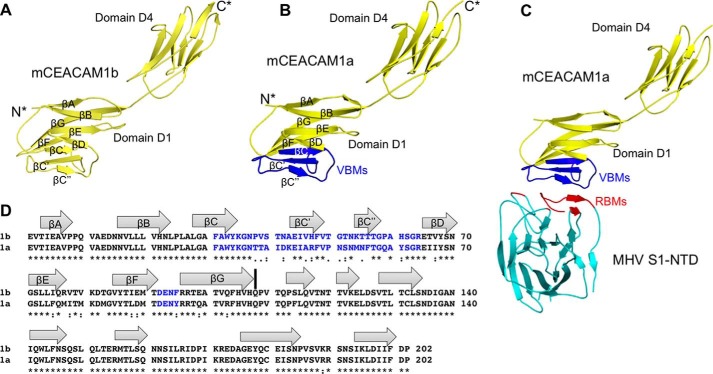
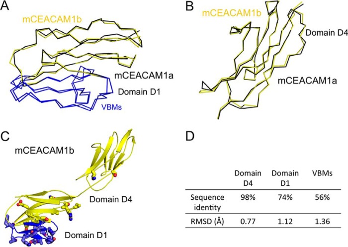
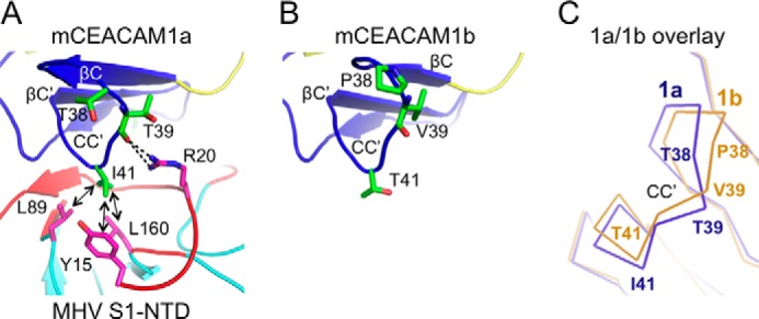
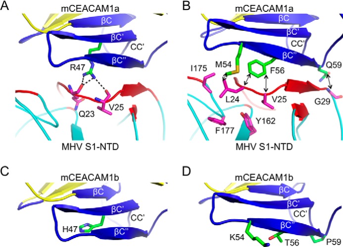
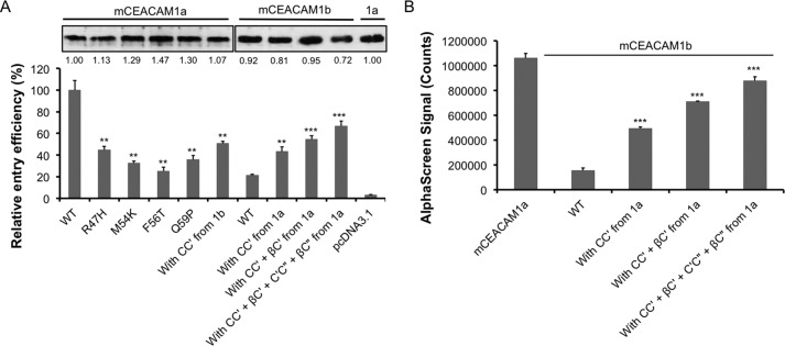
Hosts and pathogens are locked in an evolutionary arms race. To infect mice, mouse hepatitis coronavirus (MHV) has evolved to recognize mouse CEACAM1a (mCEACAM1a) as its receptor. To elude MHV infections, mice may have evolved a variant allele from the Ceacam1a gene, called Ceacam1b, producing mCEACAM1b, which is a much poorer MHV receptor than mCEACAM1a. Previous studies showed that sequence differences between mCEACAM1a and mCEACAM1b in a critical MHV-binding CC' loop partially account for the low receptor activity of mCEACAM1b, but detailed structural and molecular mechanisms for the differential MHV receptor activities of mCEACAM1a and mCEACAM1b remained elusive. Here we have determined the crystal structure of mCEACAM1b and identified the structural differences and additional residue differences between mCEACAM1a and mCEACAM1b that affect MHV binding and entry. These differences include conformational alterations of the CC' loop as well as residue variations in other MHV-binding regions, including β-strands C' and C'' and loop C'C''. Using pseudovirus entry and protein-protein binding assays, we show that substituting the structural and residue features from mCEACAM1b into mCEACAM1a reduced the viral receptor activity of mCEACAM1a, whereas substituting the reverse changes from mCEACAM1a into mCEACAM1b increased the viral receptor activity of mCEACAM1b. These results elucidate the detailed molecular mechanism for how mice may have kept pace in the evolutionary arms race with MHV by undergoing structural and residue changes in the MHV receptor, providing insight into this possible example of pathogen-driven evolution of a host receptor protein.

Keywords: X-ray crystallography; evolution; receptor structure-function; virus; virus entry.


Conflict of interest statement

The authors declare that they have no conflicts of interest with the contents of this article

Figures

FIGURE 1.
FIGURE 1.
Overall structure of mouse CEACAM1b. A, crystal structure of mCEACAM1b[D1,D4] containing the D1 and D4 domains. Secondary structures in domain D1 of mCEACAM1b[D1,D4] are named after the mCEACAM1a[D1,D4] structure (23). B, crystal structure of mCEACAM1a[D1,D4] (PDB 1L67) (23). VBMs are in blue. C, crystal structure of mCEACAM1a[D1,D4] complexed with MHV S1-NTD (PDB 3R4D) (16). MHV S1-NTD is in cyan, with receptor-binding motifs (RBMs) in red. D, sequence alignment of mCEACAM1b[D1,D4] and mCEACAM1a[D1,D4]. β-strands are shown as arrows. VBMs in mCEACAM1a and the corresponding regions in mCEACAM1b are in blue. Asterisks indicate positions that have fully conserved residues; colons indicate positions that have strongly conserved residues; periods indicate positions that have weakly conserved residues. The boundary between domains D1 and D4 is indicated by a black line. Sequence alignment was done using ClustalW (48). Structural illustrations were done using PyMOL (49).
FIGURE 2.
FIGURE 2.
Structural comparisons of mCEACAM1a and mCEACA1b. A, overlay of mCEACAM1a and mCEACAM1b in domain D1. mCEACAM1a is in black, and mCEACAM1b in yellow. VBMs in mCEACAM1a and the corresponding regions in mCEACAM1b are in blue. B, overlay of mCEACAM1a and mCEACAM1b in domain D4. C, structure of mCEACAM1b showing distribution of the residues that differ between mCEACAM1a and mCEACAM1b. Regions corresponding to VBMs in mCEACAM1a are in blue. Residues that differ between mCEACAM1a and mCEACAM1b are shown as balls and sticks. D, sequence identities and r.m.s.d. between mCEACAM1a and mCEACAM1b in different regions. See Fig. 1D for the residue ranges of domains and VBMs. r.m.s.d. were calculated using Coot (50).
FIGURE 3.
FIGURE 3.
Structural comparisons of mCEACAM1a and mCEACAM1b in MHV-binding loop CC′. A, interactions between MHV S1-NTD (from MHV strain A59) and loop CC′ of mCEACAM1a. mCEACAM1 residues are in green, and S1-NTD residues are in magenta. Hydrophobic interactions are indicated as arrows, and hydrogen bonds are indicated as dotted lines. B, structure of loop CC′ of mCEACAM1b. C, overlay of the CC′ loops from mCEACAM1a and mCEACAM1b.
FIGURE 4.
FIGURE 4.
Structural comparisons of mCEACAM1a and mCEACAM1b in MHV-binding strands βC′ and βC′′. A, interactions between MHV S1-NTD and Arg-47 in strand βC′ of mCEACAM1a. B, interactions between MHV S1-NTD and Met-54, Phe-56, and Asn-59 in strand βC′′ of mCEACAM1a. C, conformation of the side chain of His-47 in strand βC′ of mCEACAM1b. D, conformations of the side chains of Lys-54, Thr-56, and Pro-59 in strand βC′′ of mCEACAM1b.
FIGURE 5.
FIGURE 5.
Structure-guided mutational and functional characterizations of mCEACAM1a and mCEACAM1b. A, pseudovirus entry efficiency. Lentiviruses pseudotyped with MHV spike protein (from MHV strain A59) were used to enter HEK293T cells expressing mCEACAM1a or mCEACAM1b (wild type or mutant). The relative expression of each receptor was used to normalize pseudovirus entry efficiency. The pseudovirus entry mediated by wild-type mCEACAM1a was taken as 100%. Error bars indicate S.E. (n = 3). Comparisons between wild-type mCEACAM1a and mutant mCEACAM1a or between wild-type mCEACAM1b and mutant mCEACAM1b in their capabilities to mediate pseudovirus entry were done using a two-tailed t test (**, p < 0.01; ***, p < 0.001). The mutant mCEACAM1a molecules contain single mutations R47H, M54K, F56T, Q59P, or loop CC′ from mCEACAM1b (residues 38–43). The mutant mCEACAM1b molecules contain loop CC′ from mCEACAM1a (residues 38–43), loop CC′ and strand βC′ from mCEACAM1a (residues 38–51) or loop CC′, strand βC′, loop C′C′′, and strand βC′′ from mCEACAM1a (residues 38–59). B, protein-protein binding assay. The interactions between MHV S1-NTD and mCEACAM1a or mCEACAM1b (wild type or mutant) were measured using AlphaScreen assay. MHV S1-NTD with a C-terminal His6 tag and mCEAMCAM1a or mCEACAM1b (wild type or mutant) with a C-terminal human IgG4 Fc tag were attached to AlphaScreen Nickel Chelate Donor Beads and Alpha Screen protein A acceptor beads, respectively. Error bars indicate S.E. (n = 3). Comparisons between wild-type mCEACAM1b and mutant mCEACAM1b in their binding affinity for MHV S1-NTD were done using two-tailed t test (***, p < 0.001).
FIGURE 6.
FIGURE 6.
Proposed evolutionary arms race between MHV and mice. The race includes both MHV-driven evolution of mice (top) and mouse-adapting evolution of MHV (bottom). For a detailed explanation, see the “Discussion” section.

References

    1. Dawkins R., and Krebs J. R. (1979) Arms races between and within species. Proc. R. Soc. Lond. B Biol. Sci. 205, 489–511 - PubMed
    1. Van Valen L. (1973) A new evolutionary law. Evolutionary Theory 1, 1–30
    1. Perlman S., and Netland J. (2009) Coronaviruses post-SARS: update on replication and pathogenesis. Nat. Rev. Microbiol. 7, 439–450 - PMC - PubMed
    1. Li W., Wong S. K., Li F., Kuhn J. H., Huang I. C., Choe H., and Farzan M. (2006) Animal origins of the severe acute respiratory syndrome coronavirus: insight from ACE2-S-protein interactions. J. Virol. 80, 4211–4219 - PMC - PubMed
    1. Li F. (2015) Receptor recognition mechanisms of coronaviruses: a decade of structural studies. J. Virol. 89, 1954–1964 - PMC - PubMed

Publication types

LinkOut - more resources