Review
. 2017 Jan 1;28(1):1-8.
doi: 10.1091/mbc.E15-11-0769.

Of microtubules and memory: implications for microtubule dynamics in dendrites and spines

Affiliations
Review

Of microtubules and memory: implications for microtubule dynamics in dendrites and spines

Erik W Dent. Mol Biol Cell. .

Abstract

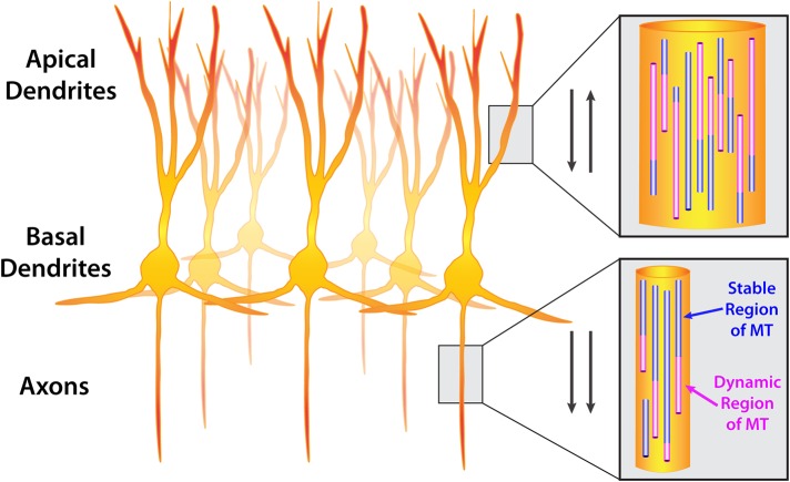
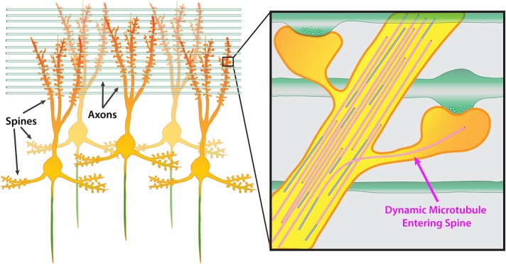
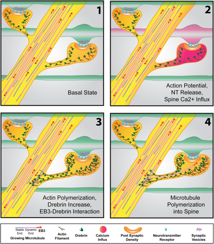
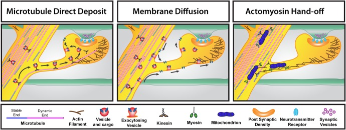
Microtubules (MTs) are cytoskeletal polymers composed of repeating subunits of tubulin that are ubiquitously expressed in eukaryotic cells. They undergo a stochastic process of polymerization and depolymerization from their plus ends termed dynamic instability. MT dynamics is an ongoing process in all cell types and has been the target for the development of several useful anticancer drugs, which compromise rapidly dividing cells. Recent studies also suggest that MT dynamics may be particularly important in neurons, which develop a highly polarized morphology, consisting of a single axon and multiple dendrites that persist throughout adulthood. MTs are especially dynamic in dendrites and have recently been shown to polymerize directly into dendritic spines, the postsynaptic compartment of excitatory neurons in the CNS. These transient polymerization events into dendritic spines have been demonstrated to play important roles in synaptic plasticity in cultured neurons. Recent studies also suggest that MT dynamics in the adult brain function in the essential process of learning and memory and may be compromised in degenerative diseases, such as Alzheimer's disease. This raises the possibility of targeting MT dynamics in the design of new therapeutic agents.


Figures

FIGURE 1:
FIGURE 1:
Orientation of microtubules in CNS neurons. A group of neurons (cortical or hippocampal) showing apical dendrites, basal dendrites, and axons. Insets show microtubule orientation in dendrites and axons. Microtubules are composed of stable (purple) and dynamic (pink) regions. Dynamic regions undergo polymerization and depolymerization, termed dynamic instability. Arrows indicate that microtubules are oriented antiparallel in dendrites (plus and minus ends distal) and parallel in axons (plus ends distal).
FIGURE 2:
FIGURE 2:
Microtubules are capable of polymerizing into dendritic spines. A group of mature CNS neurons showing dendritic spines located along both apical and basal dendrites. The axon is colored green. Green horizontal rods indicate axons from other neurons growing perpendicular to apical dendrites. Inset shows two spines along a dendrite synapsing onto two perpendicular axons. Presynaptic vesicles are shown in axons. In this example, one dynamic microtubule polymerizes into the right dendritic spine, extending well into the head of the spine.
FIGURE 3:
FIGURE 3:
Events resulting in microtubule polymerization into a dendritic spine. (1) In the basal state, microtubules, tipped by “comets” of +TIP proteins (EB3 shown here), actively polymerize throughout the dendrite (as well as the axon; not shown). Actin and the actin-binding protein drebrin are concentrated in spines. (2) As the action potential makes its way down an axon (red in second frame), resulting in neurotransmitter release at the synapse, calcium influx in the postsynaptic spine occurs. (3) Within seconds to minutes after calcium spikes in the spine, actin polymerization occurs in the spine head and neck and can extend into the dendritic shaft. The spine head increases in size due to the increased actin polymerization. Increased actin also concentrates drebrin, which interacts with EB3 protein at the tips of polymerizing microtubules in the vicinity of the spine. (4) This drebrin–EB3 interaction results in the increased probability that the polymerizing microtubule will enter the spine (on the right). The microtubule can extend well into the spine head, often to the postsynaptic density.
FIGURE 4:
FIGURE 4:
Three different routes for transporting material into dendritic spines. Left, the microtubule direct deposit model. During the time that a microtubule has polymerized into a dendritic spine, kinesin-based transport of vesicles containing cargo enter the spine along the microtubule. Release of the motor from the microtubule leads to exocytosis of the vesicle and cargo in the spine head. Middle, the membrane diffusion model. Kinesin-based transport results in movement of vesicles and cargo in the dendrite. Vesicles exocytose in the dendrite shaft and spine and diffuse in the plane of the membrane throughout the dendritic spine. Right, the actomyosin-based hand-off model of transport into spines. In this example, mitochondria are transported throughout the dendrite via kinesin-based transport but also have myosin motors attached. A hand-off occurs between kinesin–microtubule transport to actin-myosin–based transport, resulting in transport of mitochondria into the dendritic spine. Endoplasmic reticulum is transported into spines via this mechanism as well.

References

    1. Akhmanova A, Steinmetz MO. Control of microtubule organization and dynamics: two ends in the limelight. Nat Rev Mol Cell Biol. 2015;16:711–726. - PubMed
    1. Andrieux A, Salin P, Schweitzer A, Begou M, Pachoud B, Brun P, Gory-Faure S, Kujala P, Suaud-Chagny MF, Hofle G, Job D. Microtubule stabilizer ameliorates synaptic function and behavior in a mouse model for schizophrenia. Biol Psychiatry. 2006;60:1224–1230. - PubMed
    1. Baas PW, Ahmad FJ. The plus ends of stable microtubules are the exclusive nucleating structures for microtubules in the axon. J Cell Biol. 1992;116:1231–1241. - PMC - PubMed
    1. Baas PW, Ahmad FJ. Beyond taxol: microtubule-based treatment of disease and injury of the nervous system. Brain. 2013;136:2937–2951. - PMC - PubMed
    1. Baas PW, Deitch JS, Black MM, Banker GA. Polarity orientation of microtubules in hippocampal neurons: uniformity in the axon and nonuniformity in the dendrite. Proc Natl Acad Sci USA. 1988;85:8335–8339. - PMC - PubMed

Publication types