

Regulation of Neuronal Activity in Hypothalamic Vasopressin Neurons
- PMID: 28035187
- PMCID: PMC5189920
- DOI: 10.4036/iis.2015.B.07
Regulation of Neuronal Activity in Hypothalamic Vasopressin Neurons
Abstract
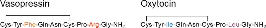
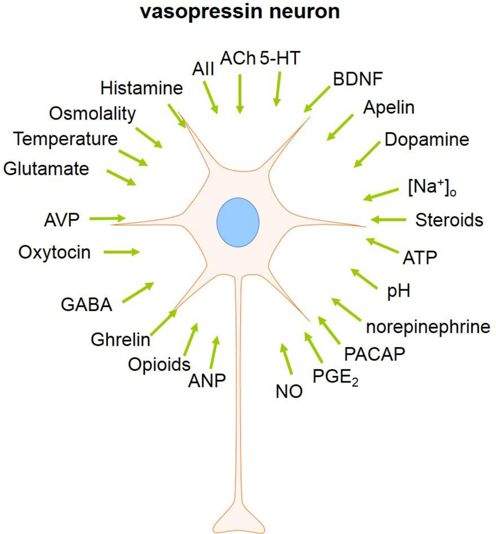
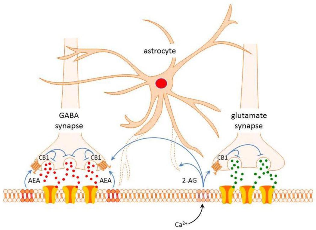
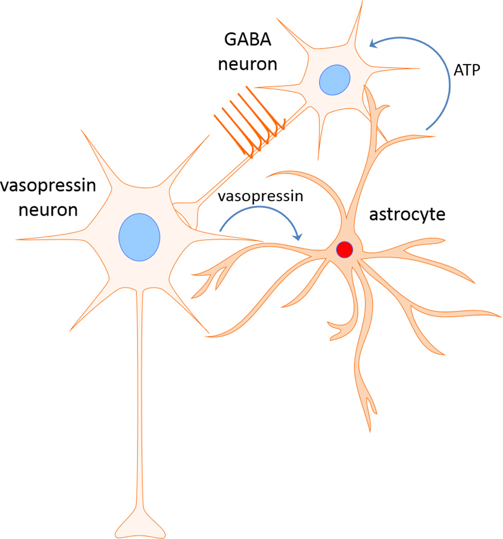
Vasopressin is a peptide hormone secreted from the posterior pituitary gland in response to various physiological and/or pathological stimuli, including changes in body fluid volume and osmolality and stress exposure. Vasopressin secretion is controlled by the electrical activity of the vasopressinergic magnocellular neurosecretory cells located in the hypothalamic supraoptic nucleus and paraventricular nucleus. Vasopressin release can occur somatodendritically in the hypothalamus or at the level of pituitary axon terminals. The electrical activity of the vasopressin neurons assumes specific patterns of electrical discharge that are under the control of several factors, including the intrinsic properties of the neuronal membrane and synaptic and hormonal inputs. It is increasingly clear that glial cells perform critical signaling functions that contribute to signal transmission in neural circuits. Astrocytes contribute to neuronal signaling by regulating synaptic and extrasynaptic neurotransmission, as well as by mediating bidirectional neuronal-glial transmission. We recently discovered a novel form of neuronal-glial signaling that exploits the full spatial domain of astrocytes to transmit dendritic retrograde signals from vasopressin neurons to distal upstream neuronal targets. This retrograde trans-neuronal-glial transmission allows the vasopressin neurons to regulate their synaptic inputs by controlling upstream presynaptic neuron firing, thus providing a powerful means of controlling hormonal output.
Keywords: astrocyte; glia; hypothalamus; neuron; paraventricular; supraoptic; synapse; vasopressin.
Figures

References
-
- Brownstein MJ, Russell JT, Gainer H. Synthesis, transport, and release of posterior pituitary hormones. Science. 1980;207:373–378. - PubMed
-
- Ludwig M, Sabatier N, Bull PM, Landgraf R, Dayanithi G, Leng G. Intracellular calcium stores regulate activity-dependent neuropeptide release from dendrites. Nature. 2002;418:85–89. - PubMed
-
- Tribollet E, Barberis C, Dubois-Dauphin M, Dreifuss JJ. Localization and characterization of binding sites for vasopressin and oxytocin in the brain of the guinea pig. Brain Res. 1992;589:15–23. - PubMed
-
- Ostrowski NL, Lolait SJ, Young WS., 3rd Cellular localization of vasopressin V1a receptor messenger ribonucleic acid in adult male rat brain, pineal, and brain vasculature. Endocrinology. 1994;135:1511–1528. - PubMed
Grants and funding
LinkOut - more resources
Full Text Sources