Review
. 2016 Dec 28;18(1):47.
doi: 10.3390/ijms18010047.

Subcellular Trafficking of Mammalian Lysosomal Proteins: An Extended View

Affiliations
Review

Subcellular Trafficking of Mammalian Lysosomal Proteins: An Extended View

Catherine Staudt et al. Int J Mol Sci. .

Abstract

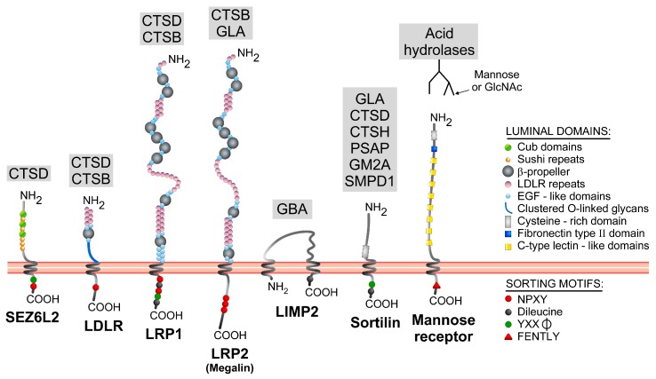
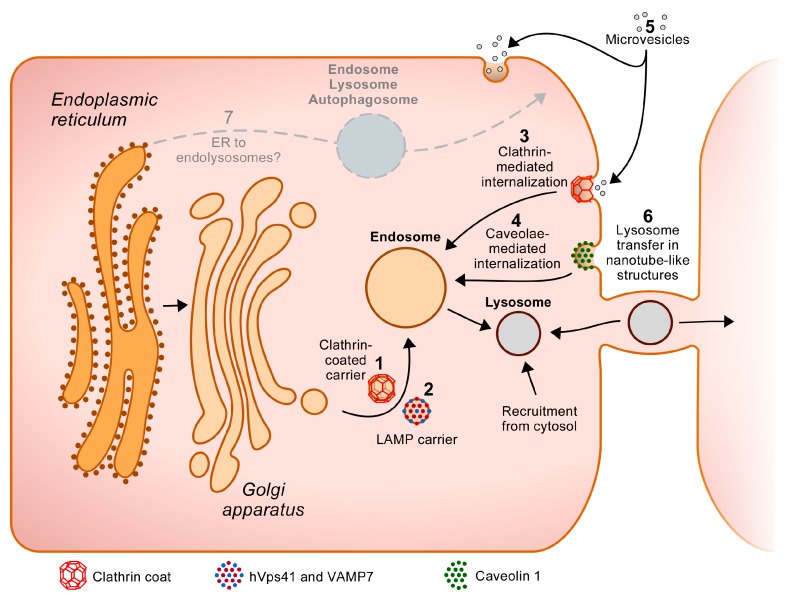
Lysosomes clear macromolecules, maintain nutrient and cholesterol homeostasis, participate in tissue repair, and in many other cellular functions. To assume these tasks, lysosomes rely on their large arsenal of acid hydrolases, transmembrane proteins and membrane-associated proteins. It is therefore imperative that, post-synthesis, these proteins are specifically recognized as lysosomal components and are correctly sorted to this organelle through the endosomes. Lysosomal transmembrane proteins contain consensus motifs in their cytosolic regions (tyrosine- or dileucine-based) that serve as sorting signals to the endosomes, whereas most lysosomal acid hydrolases acquire mannose 6-phosphate (Man-6-P) moieties that mediate binding to two membrane receptors with endosomal sorting motifs in their cytosolic tails. These tyrosine- and dileucine-based motifs are tickets for boarding in clathrin-coated carriers that transport their cargo from the trans-Golgi network and plasma membrane to the endosomes. However, increasing evidence points to additional mechanisms participating in the biogenesis of lysosomes. In some cell types, for example, there are alternatives to the Man-6-P receptors for the transport of some acid hydrolases. In addition, several "non-consensus" sorting motifs have been identified, and atypical transport routes to endolysosomes have been brought to light. These "unconventional" or "less known" transport mechanisms are the focus of this review.

Keywords: alternative receptor; lysosome; mannose 6-phosphate; sorting motif; trafficking; unconventional.


Conflict of interest statement

The authors declare no conflict of interest.

Figures

Figure 1
Figure 1
Structure and lysosomal cargo(es) of Man-6-P-alternative receptors. These receptors are mostly type I transmembrane proteins, with the exception of LIMP2, which is type III transmembrane protein. Luminal domains/repeats, and known cytosolic sorting signals are indicated on the scheme. The lysosomal proteins that bind to these receptors are listed in the grey boxes. CTSB: cathepsin B; CTSD: cathepsin D; CTSH: cathepsin H; GLA: α-galactosidase; GBA: β-glucocerebrosidase; PSAP: prosaposin; SMPD1, acid sphingomyelinase; GM2A: Ganglioside GM2 activator protein. The mannose receptor recognizes many acid hydrolases that bear terminal mannose or N-acetyl-d-glucosamine on their N-linked glycans. All three members of the LDLR family (LDLR, LRP1 and LRP2/Megalin) contain three types of domains in their N-terminal regions: β-propeller, EGF-like repeats and LDLR repeats. The latter are often involved in ligand binding. However, LRP1 binds to cathepsin D (CTSD) through amino acids 349–394 of its β-chain (last 85 kDa of the protein), i.e., a region that contains EGF-like repeats [108]. LIMP2 and β-glucocerebrosidase associate via several hydrophobic helical interfaces located on both proteins [109]. The cysteine-rich domain of sortilin has been involved in binding to several non-lysosomal ligands [110], whereas both the cysteine-rich domain and C-type lectin-like domains 4 and 5 of the mannose receptor serve as ligand binding sites [106]. SEZ6L2 contains Cub domains, which are known to mediate protein–protein interactions.
Figure 2
Figure 2
Sorting pathways to the endolysosomes. Newly synthesized lysosomal proteins are transported to the Golgi apparatus from where they reach the endosomes and, subsequently the lysosomes. One of the main transport pathways followed by these proteins is a direct route from the TGN to the endosomes (1). The lysosomal transmembrane proteins and acid hydrolase receptors (MPRs, sortilin, SEZ6L2) that are bound to their ligands are packaged at the TGN in clathrin-coated carriers that travel toward the endosomes. This packaging is driven by the binding of typical cytosolic tyrosine- or dileucine-based sorting signals, or of atypical motifs, to clathrin adaptor proteins (directly or through some other proteins). Alternatively, some transmembrane proteins (including LAMP1 and LAMP2) can be transported directly from the TGN to the endosomes in non-clathrin-coated vesicles (“LAMP carriers”) that are positive for hVps41 and VAMP7 (2). Lysosomal proteins that are not packaged in transport vesicles at the TGN, and escape to the cell surface as a consequence, can be recaptured by clathrin-mediated internalization, which is also based on the recognition of cytosolic sorting motifs by PM clathrin adaptor proteins (3). In addition, LAMP3 has been found to enter the endolysosomal system by caveolin-mediated internalization (4). Of note, some lysosomal proteins may piggyback on others to enter some of these pathways (as indicated in Table 1). The cells can also acquire some lysosomal proteins from other cells, e.g., through the capture of microvesicles by clathrin-dependent and -independent mechanisms (5) or subsequently to the transfer of exogenous lysosomes through nanotube-like structures tunneling between some cells (6). Lastly, some proportion of lysosomal proteins could, hypothetically, bypass the Golgi apparatus to reach endolysosomes or autophagosomes (which could then fuse with lysosomes) directly from the ER, or after sorting from the ER to the PM and subsequent internalization from this site (7).

Similar articles

Cited by

References

    1. De Duve C., Pressman B.C., Gianetto R., Wattiaux R., Appelmans F. Tissue fractionation studies. 6. Intracellular distribution patterns of enzymes in rat-liver tissue. Biochem. J. 1955;60:604–617. doi: 10.1042/bj0600604. - DOI - PMC - PubMed
    1. Sabatini D.D., Adesnik M. Christian de Duve: Explorer of the cell who discovered new organelles by using a centrifuge. Proc. Natl. Acad. Sci. USA. 2013;110:13234–13235. doi: 10.1073/pnas.1312084110. - DOI - PMC - PubMed
    1. Schröder B., Wrocklage C., Pan C., Jäger R., Kösters B., Schäfer H., Elsässer H.-P., Mann M., Hasilik A. Integral and associated lysosomal membrane proteins. Traffic (Cph. Den.) 2007;8:1676–1686. doi: 10.1111/j.1600-0854.2007.00643.x. - DOI - PubMed
    1. Callahan J.W., Bagshaw R.D., Mahuran D.J. The integral membrane of lysosomes: Its proteins and their roles in disease. J. Proteom. 2009;72:23–33. doi: 10.1016/j.jprot.2008.11.007. - DOI - PubMed
    1. Della Valle M.C., Sleat D.E., Zheng H., Moore D.F., Jadot M., Lobel P. Classification of subcellular location by comparative proteomic analysis of native and density-shifted lysosomes. Mol. Cell. Proteom. MCP. 2011;10:M110.006403. doi: 10.1074/mcp.M110.006403. - DOI - PMC - PubMed

LinkOut - more resources