. 2017 Jan 17;8(3):5371-5381.
doi: 10.18632/oncotarget.14260.

Combining vasculature disrupting agent and Toll-like receptor 7/8 agonist for cancer therapy

Affiliations

Combining vasculature disrupting agent and Toll-like receptor 7/8 agonist for cancer therapy

Anushree Seth et al. Oncotarget. .

Abstract

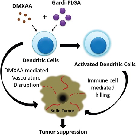
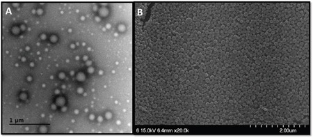
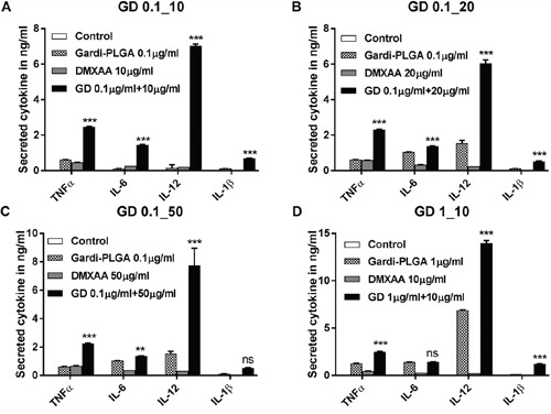
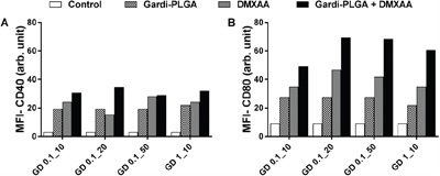
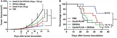
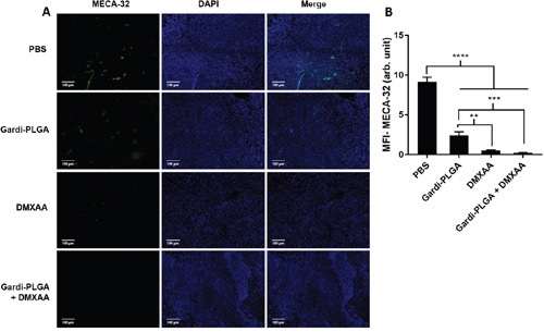
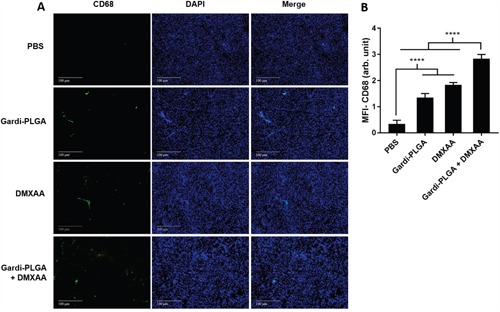
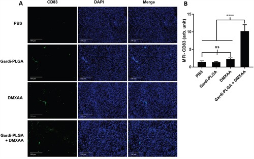
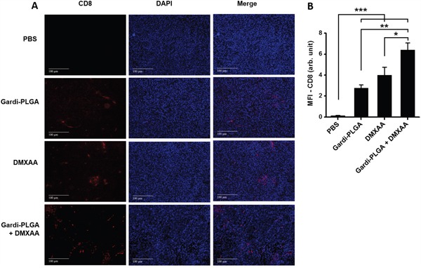
This study evaluates the effect of combination of two different treatment regimens for solid tumor therapy: vasculature targeting agent and immune-stimulation. Poly lactide-co-glycolide (PLGA) nanoparticles were synthesized for intracellular delivery of Toll-like receptor (TLR) 7/8 agonist-gardiquimod. Spherical and mono-disperse gardiquimod encapsulated PLGA nanoparticles (Gardi-PLGA), approximately 194 nm in size were formulated. Gardi-PLGA induced immune-stimulation, and vasculature disrupting agent (VDA)-5,6-Dimethylxanthenone-4-acetic acid (DMXAA) was used in combination to assessing the influence on bone marrow derived dendritic cells (BMDCs) and B16-F10 melanoma cells. The combination treatment significantly increased the levels of pro-inflammatory cytokines, indicating their activation in BMDCs, while melanoma cells remained viable. Further, mice melanoma model was established, and DMXAA was administered intraperitoneally and Gardi-PLGA was administered via an intra-tumoral injection. The combination treatments strategy significantly inhibited tumor growth as shown by tumor volume analysis, and the survival rate of the mice was found to be 63.6% (n = 11), after 54 days of tumor inoculation. Immunohistochemical findings of tumor sections treated with DMXAA confirmed the in vivo vasculature disruption. Thus, the inhibition of tumor growth can be attributed to the synergistic effect of immune stimulation caused by DC activation and vasculature disruption.

Keywords: TLR7/8 agonist; combination therapy; dendritic cells; nanoparticle; vasculature disruption.


Conflict of interest statement

CONFLICTS OF INTEREST

The authors declare no conflicts of interest.

Figures

Scheme 1
Scheme 1. Illustration depicting speculated mode of actions of combination of Gardi-PLGA and DMXAA
Gardiquimod encapsulating PLGA nanoparticles in combination with DMXAA is speculated to cause activation of dendritic cells which facilitates immune cell mediated killing of cancer cells. Vasculature disrupting agent DMXAA further aids in inhibition of tumor growth.
Figure 1
Figure 1
A. Transmission electron micrographs (scale bar, 1 μm), and B. scanning electron micrographs (scale bar, 2 μm) of the Gardi-PLGA nanoparticles.
Figure 2
Figure 2. Secretion levels of cytokine (TNFα, IL-6, IL-12 and IL-1β) by BMDCs after treatment with Gardi-PLGA, DMXAA, and their combination at various concentrations
A. Gardi-PLGA 0.1 μg/mL + DMXAA 10 μg/mL, B. Gardi-PLGA 0.1 μg/mL + DMXAA 20 μg/mL, C. Gardi-PLGA 0.1 μg/mL + DMXAA 50 μg/mL, and D. Gardi-PLGA 1 μg/mL + DMXAA 10 μg/mL. ***p < 0.001 by one-way ANOVA Test, with Tukey's multiple comparison.
Figure 3
Figure 3. Mean fluorescence intensity (MFI) of A. CD40 and B. CD80 of BMDCs after treatment with Gardi-PLGA, DMXAA, and their combination at various concentrations
Figure 4
Figure 4
A. Tumor volume (mm3) with respect to day 1 until day 27 in C57BL/6 mice immunized with Gardi-PLGA, DMXAA, and their combination. Injection time points are indicated by black arrows. Significant inhibition in tumor growth was observed after treatment with combination. *p < 0.05, ***p < 0.001, ****p < 0.0001 by one-way ANOVA Test, Bonferroni's post-test, n = 8. B. Kaplan Meier survival plot of immunized mice is shown. **p = 0.0024, n = 11.
Figure 5
Figure 5
A. Immunohistochemical images of tumor sections at day 14 after tumor inoculation for Gardi-PLGA, DMXAA, and their combination labelled with (left to right) endothelial marker (MECA-32, green), nuclear stain (DAPI, blue), and merged. B. Mean fluorescence intensity (MFI) of MECA-32 (n = 3 tumor sections, average of 10 different regions for each sample). Significant decrease in presence of endothelial cell marker confirmed vasculature disruption in combination treated groups. ****p < 0.0001, ***p < 0.001, **p < 0.01 by one-way ANOVA Test, with Tukey's multiple comparison.
Figure 6
Figure 6
A. Immunohistochemical images of tumor sections at day 14 after tumor inoculation for Gardi-PLGA, DMXAA, and their combination labelled with (left to right) macrophage marker (CD68, red), nuclear stain (DAPI, blue), and merged. B. Mean fluorescence intensity (MFI) of CD68 (n = 3 tumor sections, average of 10 different regions for each sample). Significant increases in presence of macrophage marker are shown in the treated groups. ****p < 0.0001 by one-way ANOVA Test, with Tukey's multiple comparison.
Figure 7
Figure 7
A. Immunohistochemical images of tumor sections at day 14 after tumor inoculation for Gardi-PLGA, DMXAA, and their combination labelled with (left to right) dendritic cells marker (CD83, green), nuclear stain (DAPI, blue), and merged. B. Mean fluorescence intensity (MFI) of CD83 (n = 3 tumor sections, average of 10 different regions for each sample). Significant increase of dendritic cell infiltration is shown only in combination treated group. ****p < 0.0001 by one-way ANOVA Test, with Tukey's multiple comparison.
Figure 8
Figure 8
A. Immunohistochemical images of tumor sections at day 14 after tumor inoculation for various treatment groups labelled with (left to right) T-cell marker (CD8, red), nuclear stain (DAPI, blue), and merged. B. Mean fluorescence intensity (MFI) of CD8 (n = 3 tumor sections, average of 10 different regions for each sample). There was significant increases of CD8 T cell infiltration in the DMXAA- and Gardi-PLGA-treated groups compared to PBS group, and further significant increase was observed in combination treated group. *p < 0.05, **p < 0.01, and ***p < 0.001 by one-way ANOVA Test, with Tukey's multiple comparison.

References

    1. Blagosklonny MV. How Avastin potentiates chemotherapeutic drugs: action and reaction in antiangiogenic therapy. Cancer Biol Ther. 2005;4:1307–1310. - PubMed
    1. Kim R, Emi M, Tanabe K. Cancer immunoediting from immune surveillance to immune escape. Immunology. 2007;121:1–14. - PMC - PubMed
    1. Schreiber RD, Old LJ, Smyth MJ. Cancer Immunoediting: Integrating Immunity's Roles in Cancer Suppression and Promotion. Science. 2011;331:1565–1570. - PubMed
    1. Cheng M, Chen Y, Xiao W, Sun R, Tian Z. NK cell-based immunotherapy for malignant diseases. Cell Mol Immunol. 2013;10:230–252. - PMC - PubMed
    1. Palucka K, Banchereau J. Cancer immunotherapy via dendritic cells. Nat Rev Cancer. 2012;12:265–277. - PMC - PubMed

MeSH terms