. 2017 Mar 21;8(12):19403-19412.
doi: 10.18632/oncotarget.14292.

Heparanase augments insulin receptor signaling in breast carcinoma

Affiliations

Heparanase augments insulin receptor signaling in breast carcinoma

Rachel Goldberg et al. Oncotarget. .

Abstract

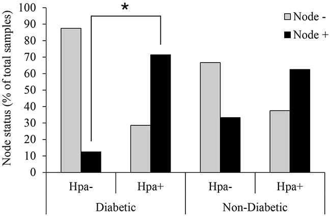
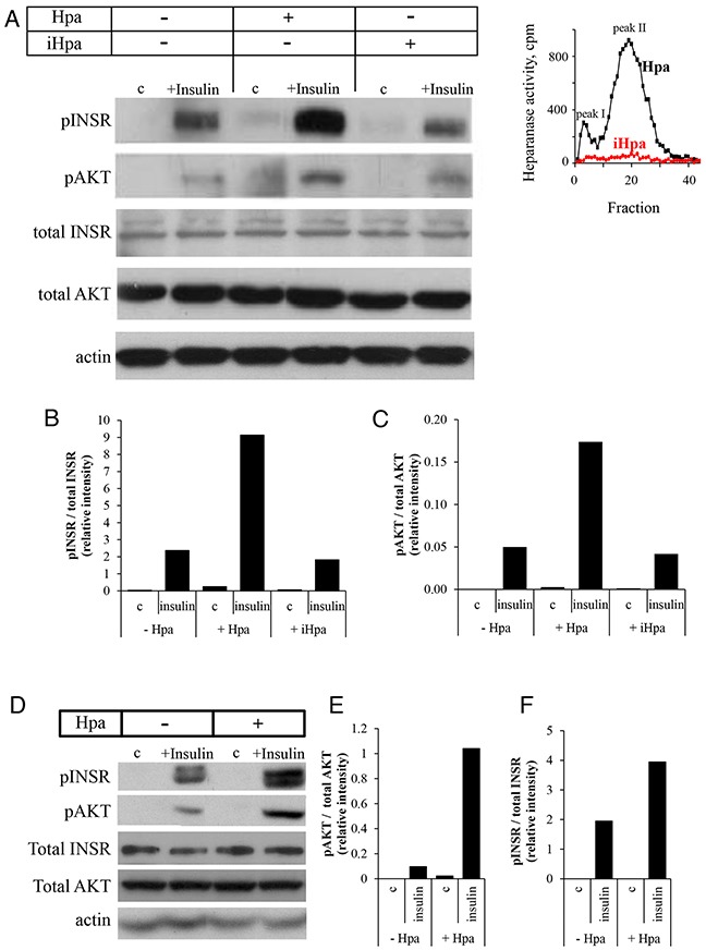
Recently, growing interest in the potential link between metabolic disorders (i.e., diabetes, obesity, metabolic syndrome) and breast cancer has mounted, including studies which indicate that diabetic/hyperinsulinemic women have a significantly higher risk of bearing breast tumors that are more aggressive and associated with higher death rates. Insulin signaling is regarded as a major contributor to this phenomenon; much less is known about the role of heparan sulfate-degrading enzyme heparanase in the link between metabolic disorders and cancer.In the present study we analyzed clinical samples of breast carcinoma derived from diabetic/non-diabetic patients, and investigated effects of heparanase on insulin signaling in breast carcinoma cell lines, as well as insulin-driven growth of breast tumor cells.We demonstrate that heparanase activity leads to enhanced insulin signaling and activation of downstream tumor-promoting pathways in breast carcinoma cells. In agreement, heparanase enhances insulin-induced proliferation of breast tumor cells in vitro. Moreover, analyzing clinical data from diabetic breast carcinoma patients, we found that concurrent presence of both diabetic state and heparanase in tumor tissue (as opposed to either condition alone) was associated with more aggressive phenotype of breast tumors in the patient cohort analyzed in our study (two-sided Fisher's exact test; p=0.04). Our findings highlight the emerging role of heparanase in powering effect of hyperinsulinemic state on breast tumorigenesis and imply that heparanase targeting, which is now under intensive development/clinical testing, could be particularly efficient in a growing fraction of breast carcinoma patients suffering from metabolic disorders.

Keywords: breast carcinoma; diabetes; heparanase; insulin receptor.


Conflict of interest statement

CONFLICTS OF INTEREST

The authors declare that they have no conflict of interest.

Figures

Figure 1
Figure 1. Heparanase expression and lymph node involvement in diabetic breast carcinoma patients
Human breast carcinoma tissue samples (biopsies) were processed for immunohistochemistry with anti-heparanase antibody (733) directed against a synthetic peptide (158KKFKNSTYRSSSVD171) corresponding to the N-terminus of the 50-kDa subunit of the heparanase enzyme, as described in [28, 57, 58]. Diabetic state, BMI and lymph node status were determined from patient history. Node+: patients with lymph node positive tumors; Node-: patients with lymph node negative tumors. Two-sided Fisher's exact test confirmed that in diabetic patients heparanase-positive tumors are more likely to spread into lymph nodes (*p=0.04). Presence of heparanase in non-diabetic breast tumor samples did not confer statistical significant difference in lymph node involvement.
Figure 2
Figure 2. Recombinant heparanase enzyme enhances insulin receptor signaling pathway in breast cancer cells
A–C. Human breast carcinoma MCF-7 cells were serum-starved overnight and then either remained untreated (c) or stimulated with insulin (100 nM) for 30 min in the absence or presence of 0.8 μg/ml active recombinant heparanase (Hpa) or heat-inactivated heparanase (iHpa). A. Cell lysates containing equivalent amounts of total protein were then immunoblotted using antibody specific for phospho-insulin receptor (pINSR), phospho-AKT (pAKT), total INSR, total AKT or total actin. B, C. The band intensity was quantified using ImageJ software; intensity ratio for pINSR/total INSR (B) and pAKT/total AKT (C) are shown. The data are representative of three independent experiments. Inset: Enzymatic activity in samples of Hpa (black line) and iHpa (red line) was examined as described in Methods. D–F. Mouse E0771 breast cancer cells (characterized by low endogenous levels of heparanase) were serum-starved overnight and then treated as described in A-C. Intensity ratio for pINSR/total INSR (E) and pAKT/total AKT (F) are shown. The data are representative of three independent experiments.
Figure 3
Figure 3. Heparanase overexpression enhances insulin receptor signaling pathway and augments proliferative response to insulin in MCF-7 cells
A. Heparanase overexpression in MCF7-Hpa (as compared to control MCF7-mock) cells was confirmed by activity assay, as described in Method and refs.(19, 54). B. MCF7-mock and MCF7-Hpa cells were serum-starved overnight and then either remained untreated (c) or stimulated with insulin (100 nM) for 15 and 30 min. Cell lysates containing equivalent amounts of total protein were then immunoblotted using antibody specific for pINSR, pAKT, total INSR, total AKT or total actin. The data shown are representative of three independent experiments. C. Presence of heparanase specific inhibitor SST0001 (10 μg/ml) reduced pINSR levels in MCF7-Hpa cells treated with insulin. D. Bar graph demonstrates the increase in proliferation of MCF7-mock and MCF7-Hpa cells cultured for 72 h in the absence (gray bars) or presence of insulin (100 nM, black bars), analyzed by MTS Cell Proliferation Assay. Note that presence of insulin does not confer statistically significant increase in proliferation of MCF7-mock cells (lacking heparanase activity). In contrast, in heparanase overexpressing MCF7-Hpa cells proliferation rate was significantly higher in the presence of insulin. *p<0.04 Two-sided Student's t test. Error bars represent ± SD. The data shown are representative of three independent experiments.

References

    1. Baumgarten SC, Frasor J. Minireview: Inflammation: an instigator of more aggressive estrogen receptor (ER) positive breast cancers. Mol Endocrinol. 2012;26:360–71. - PMC - PubMed
    1. Hursting SD. Obesity, energy balance, and cancer: a mechanistic perspective. Cancer Treat Res. 2014;159:21–33. - PubMed
    1. Khandekar MJ, Cohen P, Spiegelman BM. Molecular mechanisms of cancer development in obesity. Nat Rev Cancer. 2011;11:886–95. - PubMed
    1. Lipscombe L, Fischer H, Austin P, Fu L, Jaakkimainen RL, Ginsburg O, Rochon P, Narod S, Paszat L. The association between diabetes and breast cancer stage at diagnosis: a population-based study. Breast Cancer Research and Treatment. 2015;150:613–20. - PubMed
    1. Peairs KS, Barone BB, Snyder CF, Yeh HC, Stein KB, Derr RL, Brancati FL, Wolff AC. Diabetes Mellitus and Breast Cancer Outcomes: A Systematic Review and Meta-Analysis. Journal of Clinical Oncology. 2011;29:40–6. - PMC - PubMed