. 2017 Feb 14;8(7):11030-11041.
doi: 10.18632/oncotarget.14212.

Chromosome nondisjunction during bipolar mitoses of binucleated intermediates promote aneuploidy formation along with multipolar mitoses rather than chromosome loss in micronuclei induced by asbestos

Affiliations

Chromosome nondisjunction during bipolar mitoses of binucleated intermediates promote aneuploidy formation along with multipolar mitoses rather than chromosome loss in micronuclei induced by asbestos

Tianwei Zhang et al. Oncotarget. .

Abstract

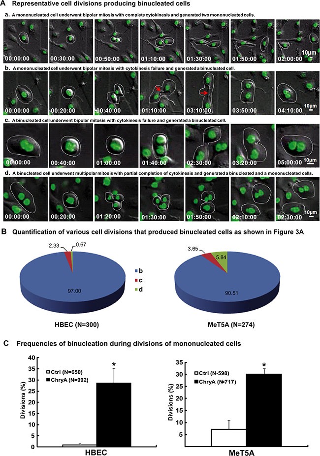
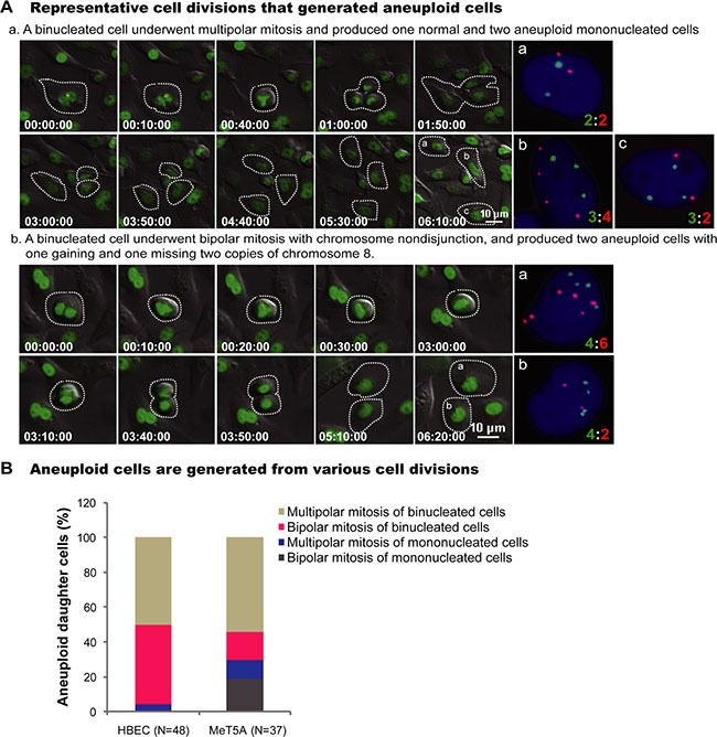
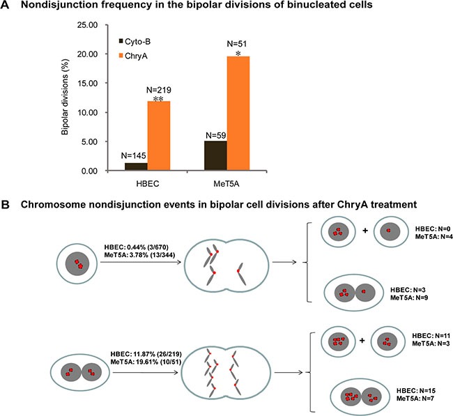
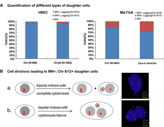
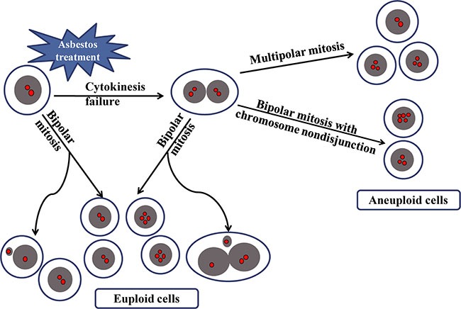
Asbestos is a well-known occupational carcinogen that can cause aneuploidy during the early stages of neoplastic development. To explore the origins of asbestos-induced aneuploidy, we performed long-term live-cell imaging followed by fluorescence in situ hybridization of chromosomes 8 and 12 in human bronchial epithelial (HBEC) and mesothelial (MeT5A) cells. We demonstrate that asbestos induces aneuploidy via binucleated intermediates resulting from cytokinesis failure. On the one hand, asbestos increases chromosome nondisjunction during bipolar divisions of binucleated intermediates and produces near-tetraploidy. On the other hand, asbestos increases multipolar divisions of binucleated intermediates to produce aneuploidy. Surprisingly, chromosomes in asbestos-induced micronucleated cells are not truly lost by the cells, and do not contribute to aneuploid cell formation in either cell type. These results clarify the cellular source of asbestos-induced aneuploidy. In particular, they show the asbestos-induced disruption of bipolar chromosomal segregation in tetraploid cells, thereby demonstrating the causality between binucleated intermediates and aneuploidy evolution, rather than chromosome loss in micronuclei.

Keywords: aneuploidy; asbestos; binucleated cell; chromosome nondisjunction; tetraploid.


Conflict of interest statement

CONFLICTS OF INTEREST

The authors declare no conflicts of interest.

Figures

Figure 1
Figure 1. Asbestos induces binucleated cells through cytokinesis failure
(A) Serial images showed representative HBEC cell normal division producing two mononucleated cells (Supplementary Movie S1) and divisions producing binucleated cells (Supplementary Movie S2–S4). Red arrows indicate asbestos across the cytoplasmic bridge region during divisions. Time is indicated in hours: minutes: seconds. (B) Quantification of various cell divisions producing binucleated daughter cells in HBEC and MeT5A cells after chrysotile treatment (N: the number of binucleated daughter cells analyzed). All the data were from at least two independent live-cell imaging experiments. (C) The frequency of binucleation in divisions was compared between untreated (Ctrl) and chrysotile-treated (ChryA) mononucleated HBEC and MeT5A cells (N: the number of divisions analyzed). *p < 0.001, 2 × 2 χ2 test.
Figure 2
Figure 2. Representative cell divisions generating aneuploid cells
(A) Serial images showed two representative HBEC cell divisions leading to aneuploid cells: (a) A binucleated cell underwent tripolar mitosis and produced three daughter cells with chromosome 12:8 compositions of 2:2, 3:4 and 3:2, respectively (Supplementary Movie S5); (b) A binucleated cell underwent bipolar mitosis with nondisjunction of chromosome 8, and produced two daughter cells with 4:6 and 4:2 for chromosome 12:8 (Supplementary Movie S6). Time is indicated in hours: minutes: seconds. The number of chromosomes in daughter cells presenting in the last frame of time-lapse imaging was directly assayed by FISH using chromosome 8 (Red) and 12 (Green) -specific probes immediately after live-cell imaging. (B) Quantification of cell divisions that produced aneuploid cells. N, the number of aneuploid daughter cells analyzed. Data were summarized from analysis of FISH signals following long-term live-cell imaging from at least two independent experiments.
Figure 3
Figure 3. Asbestos increases frequency of chromosome nondisjunction in the bipolar divisions of binucleated cells
(A) The frequency of chromosome nondisjunction was compared in the bipolar divisions of cytochalasin-B induced (Cyto-B) and chrysotile-induced (ChryA) binucleated cells (N: the number of bipolar divisions analyzed). *p < 0.05, **p < 0.001, 2 × 2 χ2 test. The number of chromosomes in daughter cells presenting in the last frame of time-lapse imaging was directly assayed by FISH using chromosome 8 (Red) and 12 (Green) -specific probes immediately after live-cell imaging from at least two independent experiments. (B) A schematic diagram summarizing the chromosome nondisjunction events in the bipolar divisions of binucleated HBEC and MeT5A cells after chrysotile treatment (N: the number of bipolar divisions analyzed).
Figure 4
Figure 4. Chromosome loss through micronuclei after chrysotile treatment does not generate aneuploid cells
(A) Quantification of different types of daughter cells in untreated and chrysotile-treated (ChryA) HBEC and MeT5A cells. (MN+) indicates micronuclei-bearing cells, (MN+; chr 8/12-) indicates micronuclei-bearing cells without chromosome 8 or 12 FISH signals in the micronuclei, while (MN+; chr 8/12+) indicates micronuclei-bearing cells with chromosome 8 or 12 FISH signals in the micronuclei (N: the number of daughter cells analyzed). (B) A schematic diagram showed two types of divisions in HBEC cells resulting in micronucleated daughter cells with chromosome 8 or 12 FISH signals in the micronuclei (MN+; chr 8/12+): (a) the chromosome was distributed into the right daughter cell along with the micronucleus; (b) daughter cells fused and formed a binucleated or multinucleated cell, both of which types generating euploid cells as shown by the representative FISH images (right). All the data were summarized from FISH following long-term live-cell imaging from at least two independent experiments.
Figure 5
Figure 5. A schematic diagram summarizing the origin of aneuploid cells induced by asbestos
Cytokinesis failure of mononucleated cellsbecause of asbestos treatment leads to binucleated cells, which produced aneuploid cells through either multipolar mitosis or bipolar mitosis with chromosome nondisjunction. While micronucleated cells resulted from bipolar mitosis of mononucleated or binucleated cells do not contribute to the generation of aneuploid cells.

References

    1. Gilham C, Rake C, Burdett G, Nicholson AG, Davison L, Franchini A, Carpenter J, Hodgson J, Darnton A, Peto J. Pleural mesothelioma and lung cancer risks in relation to occupational history and asbestos lung burden. Occup Environ Med. 2016;73:290–299. - PMC - PubMed
    1. Markowitz S. Asbestos-related lung cancer and malignant mesothelioma of the pleura: selected current issues. Semin Respir Crit Care Med. 2015;36:334–346. - PubMed
    1. Neumann V, Loseke S, Nowak D, Herth FJ, Tannapfel A. Malignant pleural mesothelioma: incidence, etiology, diagnosis, treatment, and occupational health. Dtsch Arztebl Int. 2013;110:319–326. - PMC - PubMed
    1. Prazakova S, Thomas PS, Sandrini A, Yates DH. Asbestos and the lung in the 21st century: an update. Clin Respir J. 2014;8:1–10. - PubMed
    1. Negrini S, Gorgoulis VG, Halazonetis TD. Genomic instability—an evolving hallmark of cancer. Nat Rev Mol Cell Biol. 2010;11:220–228. - PubMed

LinkOut - more resources