. 2017 Mar 1:50:437-449.
doi: 10.1016/j.actbio.2016.12.049. Epub 2016 Dec 27.

Encapsulation of primary salivary gland cells in enzymatically degradable poly(ethylene glycol) hydrogels promotes acinar cell characteristics

Affiliations

Encapsulation of primary salivary gland cells in enzymatically degradable poly(ethylene glycol) hydrogels promotes acinar cell characteristics

Andrew D Shubin et al. Acta Biomater. .

Abstract

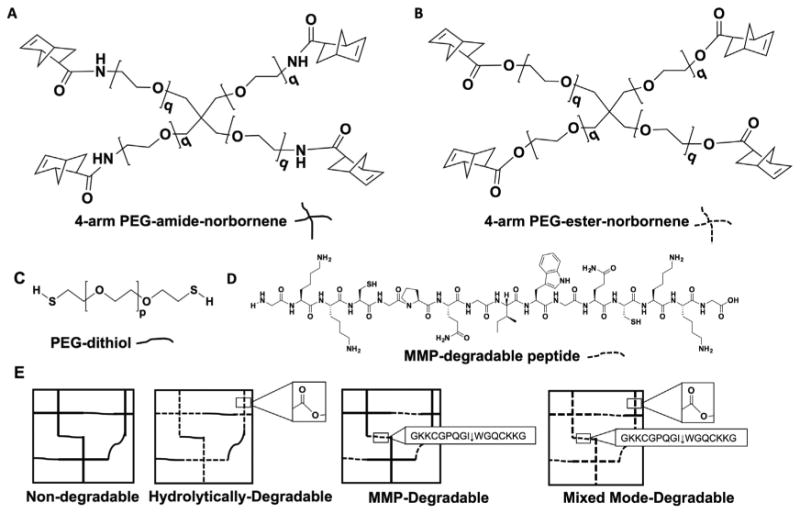
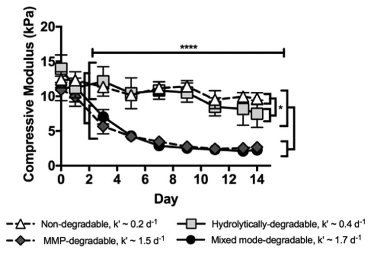
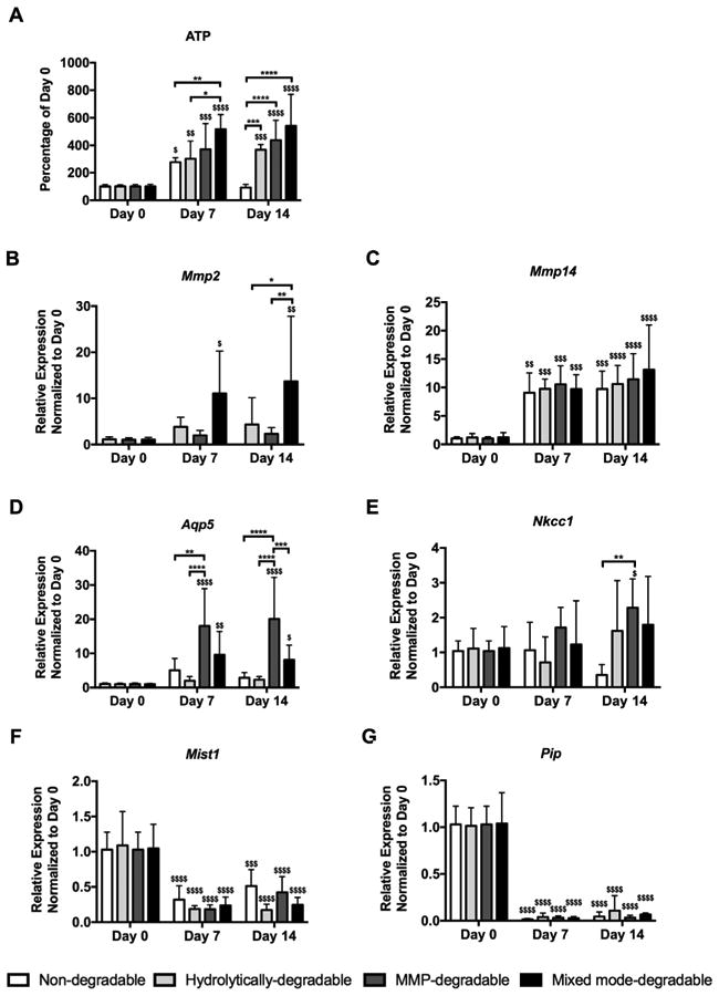
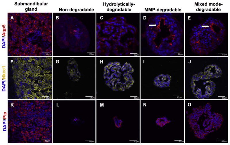
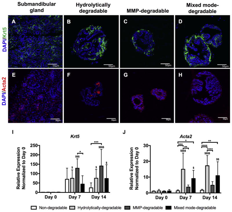
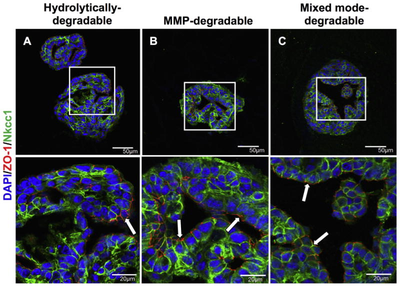
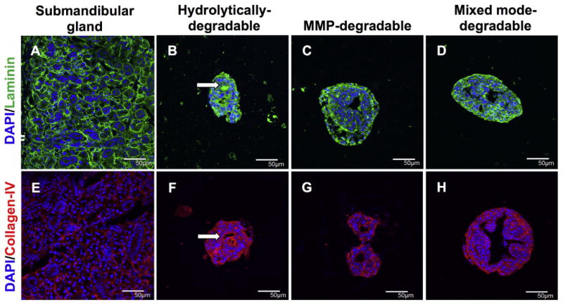
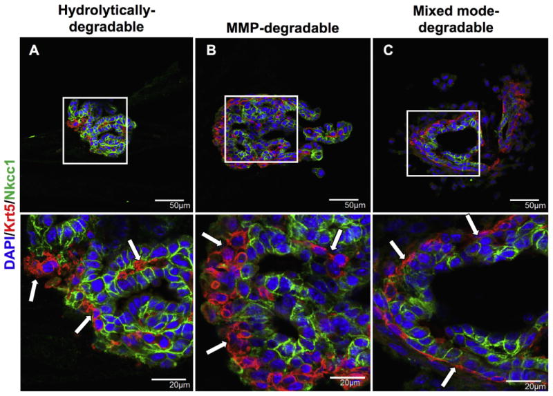
Radiation therapy for head and neck cancers leads to permanent xerostomia due to the loss of secretory acinar cells in the salivary glands. Regenerative treatments utilizing primary submandibular gland (SMG) cells show modest improvements in salivary secretory function, but there is limited evidence of salivary gland regeneration. We have recently shown that poly(ethylene glycol) (PEG) hydrogels can support the survival and proliferation of SMG cells as multicellular spheres in vitro. To further develop this approach for cell-based salivary gland regeneration, we have investigated how different modes of PEG hydrogel degradation affect the proliferation, cell-specific gene expression, and epithelial morphology within encapsulated salivary gland spheres. Comparison of non-degradable, hydrolytically-degradable, matrix metalloproteinase (MMP)-degradable, and mixed mode-degradable hydrogels showed that hydrogel degradation by any mechanism is required for significant proliferation of encapsulated cells. The expression of acinar phenotypic markers Aqp5 and Nkcc1 was increased in hydrogels that are MMP-degradable compared with other hydrogel compositions. However, expression of secretory acinar proteins Mist1 and Pip was not maintained to the same extent as phenotypic markers, suggesting changes in cell function upon encapsulation. Nevertheless, MMP- and mixed mode-degradability promoted organization of polarized cell types forming tight junctions and expression of the basement membrane proteins laminin and collagen IV within encapsulated SMG spheres. This work demonstrates that cellularly remodeled hydrogels can promote proliferation and gland-like organization by encapsulated salivary gland cells as well as maintenance of acinar cell characteristics required for regenerative approaches. Investigation is required to identify approaches to further enhance acinar secretory properties.

Statement of significance: Regenerative strategies to replace damaged salivary glands require the function and organization of acinar cells. Hydrogel-based approaches have shown promise to control cell function and phenotype. However, little is known about how specific parameters, such as the mechanism of hydrogel degradation (e.g., hydrolytic or enzymatic), influence the viability, proliferation, organization, and phenotype of salivary gland cells. In this work, it is shown that hydrogel-encapsulated primary salivary gland cell proliferation is dependent upon hydrogel degradation. Hydrogels crosslinked with enzymatically degradable peptides promoted the expression of critical acinar cell markers, which are typically downregulated in primary cultures. Furthermore, salivary gland cells encapsulated in enzymatically- but not hydrolytically-degradable hydrogels displayed highly organized and polarized salivary gland cell markers, which mimics characteristics found in native gland tissue. In sum, results indicate that salivary gland cells respond to cellularly remodeled hydrogels, resulting in self-assembly and organization akin to acini substructures of the salivary gland.

Keywords: Acinar cells; Degradation; Hydrogel; Poly(ethylene glycol); Salivary gland.


Figures

Fig. 1
Fig. 1
Macromer chemistry dictates the mode of degradation of PEG hydrogels. The composition of the four types of hydrogels used for SMG sphere encapsulations: non-degradable, hydrolytically-degradable, MMP-degradable, and mixed mode-degradable. 4-arm PEG norbornene q = 113 containing either a non-degradable amide (A) or hydrolytically-degradable ester (B). Dithiol crosslinkers used were either PEG-dithiol (p = 73, C) or the MMP-degradable peptide (D). (E) Exposure to 5 mW/cm2 365 nm light and the photoinitiator lithium phenyl-2,4,6-trimethylbenzoylphosphinate (LAP) results in thiol-ene mediated polymerization of hydrogel macromers in 3 min.
Fig. 2
Fig. 2
MMP-degradable and mixed mode-degradable hydrogels degrade upon treatment with collagenase (k′ = 1.5 and 1.7 d−1) while non-degradable and hydrolytically-degradable hydrogels exhibit modest degradation over the same time course (k′ = 0.2 and 0.4 d−1), as expected based on hydrogel composition. Degradation was measured over time using unconfined compression with 1 μg/mL collagenase treatment. N = 3–5; error bars denote ± standard deviation; * = p < 0.05, **** = p < 0.0001 when comparing hydrogels specified by brackets. Significant differences between hydrolytically-degradable hydrogels and MMP-degradable and mixed mode-degradable hydrogels at day 0 (p < 0.01) and between non-degradable hydrogels and MMP-degradable hydrogels at day 1 (p < 0.05), which are likely due to slight variability in hydrogel formulations, are not denoted due to space constraints.
Fig. 3
Fig. 3
MMP-degradable hydrogels promote SMG sphere proliferation and expression of acinar cell markers Aqp5 and Nkcc1 while secretory markers Pip and Mist1 are reduced. (A) ATP levels normalized to day 0 were used to measure proliferation of encapsulated SMG cells. Quantitative PCR was used to measure gene expression, relative to LE32 (housekeeping gene), of Mmp2 (B), Mmp14 (C), Aqp5 (D), Nkcc1 (E), Mist1 (F), and Pip (G) of SMG cell spheres at day 0 (pre-encapsulation) and days 7 and 14 (post-encapsulation). N = 5–10 per hydrogel type per time point; error bars denote ± standard deviation; compared to day 0, $ = p < 0.05, $$ = p < 0.01, $$$ = p < 0.001, $$$$ = p < 0.0001; comparing hydrogel types denoted by brackets, * = p < 0.05, ** = p < 0.01, *** = p < 0.001, **** = p < 0.0001.
Fig. 4
Fig. 4
MMP-degradability promotes the apicobasal localization of acinar cell markers in SMG cell spheres. Immunohistochemistry was used to analyze Aqp5 (red, A-E), Nkcc1 (yellow, F-J), and Pip (red, K-O) in submandibular glands (A,F,K) and SMG cell spheres encapsulated in non-degradable (B,G,L), hydrolytically-degradable (C,H,M), MMP-degradable (D,I,N), and mixed mode-degradable (E,J,O) hydrogels at day 14. Arrows indicate apical localization. DAPI counterstain was used to visualize nuclei (blue). Scale bars represent either 50 μm (A, F-O) or 20 μm (B-E). (For interpretation of the references to colour in this figure legend, the reader is referred to the web version of this article.)
Fig. 5
Fig. 5
Krt5-positive cells in degradable hydrogels are mainly ductal. Immunohistochemistry was used to analyze Krt5 (green, A-D) and Acta2 (red, E-H) in submandibular glands (A,E) and SMG cell spheres encapsulated in hydrolytically-degradable (B,F), MMP-degradable (C,G), and mixed mode-degradable (D,H) hydrogels at day 14. DAPI counterstain was used to visualize nuclei (blue). Scale bars represent 50 μm. Quantitative PCR was used to measure gene expression, relative to LE32 (housekeeping gene), of Krt5 (I) and Acta2 (J) in SMG cell spheres at day 0 (pre-encapsulation) and days 7 and 14 (post-encapsulation). N = 5–7 per hydrogel type per time point; error bars denote ± standard deviation; compared to day 0, $ = p < 0.05, $$ = p < 0.01, $$$ = p < 0.001, $$$$ = p < 0.0001; comparing hydrogel types denoted by brackets, * = p < 0.05, ** = p < 0.01, *** = p < 0.001, **** = p < 0.0001. (For interpretation of the references to colour in this figure legend, the reader is referred to the web version of this article.)
Fig. 6
Fig. 6
MMP-degradability promotes formation of tight junctions and apicobasal polarization. Immunohistochemistry was used to analyze co-stained ZO-1 (red) and Nkcc1 (green) in SMG cell spheres encapsulated in hydrolytically-degradable (A), MMP-degradable (B), and mixed mode-degradable (C) hydrogels at day 14. DAPI counterstain was used to visualize nuclei (blue) and arrows highlight areas of ZO-1 staining. Scale bars represent either 50 μm (top row) or 20 μm (bottom row). (For interpretation of the references to colour in this figure legend, the reader is referred to the web version of this article.)
Fig. 7
Fig. 7
MMP-degradability promotes basolateral localization of basement membrane proteins. Immunohistochemistry was used to analyze laminin (green, A-D) and collagen-IV (red, E-H) in submandibular glands (A,E) and SMG cell spheres encapsulated in hydrolytically-degradable (B,F), MMP-degradable, and mixed mode-degradable hydrogels (D,H) at day 14. Arrows indicate apical localization. DAPI counterstain was used to visualize nuclei (blue). Scale bars represent 50 μm. (For interpretation of the references to colour in this figure legend, the reader is referred to the web version of this article.)
Fig. 8
Fig. 8
MMP-degradability promotes the localization of Nkcc1-positive acinar cells at the lumen and Krt5-positive duct/myoepithelial cells at the periphery of SMG cell spheres. Immunohistochemistry was used to analyze co-stained Krt5 (red) and Nkcc1 (green) in hydrolytically-degradable (A), MMP-degradable (B), and mixed mode-degradable (C) hydrogels at day 14. DAPI counterstain was used to visualize nuclei (blue) and arrows are used to accentuate the organizational differences in Krt5 staining. Scale bars represent either 50 μm (top row) or 20 μm (bottom row). (For interpretation of the references to colour in this figure legend, the reader is referred to the web version of this article.)

Similar articles

Cited by

References

    1. Jensen SB, Pedersen AM, Vissink A, Andersen E, Brown CG, Davies AN, Dutilh J, Fulton JS, Jankovic L, Lopes NN, Mello AL, Muniz LV, Murdoch-Kinch CA, Nair RG, Napenas JJ, Nogueira-Rodrigues A, Saunders D, Stirling B, Von Bultzingslowen I, Weikel DS, Elting LS, Spijkervet FK, Brennan MT, Salivary S. Gland Hypofunction/Xerostomia, G. Oral Care Study, O. Multinational Association of Supportive Care in Cancer /International Society of Oral, A systematic review of salivary gland hypofunction and xerostomia induced by cancer therapies: management strategies and economic impact. Support Care Cancer. 2010;18:1061–1079. - PubMed
    1. Vissink A, Jansma J, Spijkervet FK, Burlage FR, Coppes RP. Oral sequelae of head and neck radiotherapy. Crit Rev Oral Biol Med. 2003;14:199–212. - PubMed
    1. Arany S, Benoit DS, Dewhurst S, Ovitt CE. Nanoparticle-mediated gene silencing confers radioprotection to salivary glands in vivo. Mol Ther. 2013;21:1182–1194. - PMC - PubMed
    1. Arany S, Xu Q, Hernady E, Benoit DS, Dewhurst S, Ovitt CE. Pro-apoptotic gene knockdown mediated by nanocomplexed siRNA reduces radiation damage in primary salivary gland cultures. J Cell Biochem. 2012;113:1955–1965. - PMC - PubMed
    1. Cotrim AP, Sowers A, Mitchell JB, Baum BJ. Prevention of irradiation-induced salivary hypofunction by microvessel protection in mouse salivary glands. Mol Ther. 2007;15:2101–2106. - PubMed