

Estrogen levels influence medullary bone quantity and density in female house finches and pine siskins
- PMID: 28039066
- PMCID: PMC5392170
- DOI: 10.1016/j.ygcen.2016.12.015
Estrogen levels influence medullary bone quantity and density in female house finches and pine siskins
Abstract
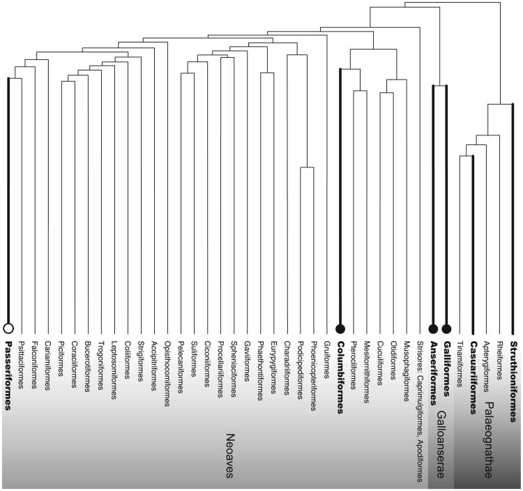
Medullary bone, a non-structural osseous tissue, serves as a temporary storage site for calcium that is needed for eggshell production in a number of avian species. Previous research focusing primarily on domesticated species belonging to the Anseriformes, Galliformes, and Columbiformes has indicated that rising estrogen levels are a key signal stimulating medullary bone formation; Passeriformes (which constitute over half of extant bird species and are generally small) have received little attention. In the current study, we examined the influence of estrogen on medullary bone and cortical bone in two species of Passeriformes: the Pine Siskin (Spinus pinus) and the House Finch (Haemorhous mexicanus). Females of these species received either an estradiol implant or were untreated as a control. After 4.5-5months, reproductive condition was assessed and leg (femora) and wing (humeri) bones were collected for analysis using high-resolution (10μm) micro-computed tomography scanning. We found that in both species estradiol-treated females had significantly greater medullary bone quantity in comparison to untreated females, but we found no differences in cortical bone quantity or microarchitecture. We were also able to examine medullary bone density in the pine siskins and found that estradiol treatment significantly increased medullary bone density. Furthermore, beyond the effect of the estradiol treatment, we observed a relationship between medullary bone quantity and ovarian condition that suggests that the timing of medullary bone formation may be related to the onset of yolk deposition in these species. Further research is needed to better understand the precise timing and endocrine regulation of medullary bone formation in Passerines and to determine the extent to which female Passerines rely on medullary bone calcium during the formation of calcified eggshells.
Keywords: Estradiol treatment; Medullary bone; Microcomputed tomography.
Copyright © 2016 Elsevier Inc. All rights reserved.
Figures


References
-
- Acenzi A, Francois C, Bocciarelli D. On the bone induced by estrogens in birds. J Ultrastruct Res. 1963;8:491–505. - PubMed
-
- Ankney CD, MacInnes CD. Nutrient reserves and reproductive performance of female lesser snow geese. Auk. 1978;95:459–471.
-
- Ankney CD, Scott DM. Changes in nutrient reserves and diet of breeding brown-headed cowbirds. Auk. 1980;97:684–696.
-
- Bloom W, Bloom MA, McLeah FC. Calcification and ossification. Medullary bone changes in the reproductive cycle of female pigeons. Anat Rec. 1941;81:443–475.
-
- Bloom MA, McLean FC, Bloom W. Calcification and ossification. The formation of medullary bone in male and castrate pigeons under the influence of sex hormones. Anat Rec. 1942;83:99–120.
Publication types
MeSH terms
Substances
Grants and funding
LinkOut - more resources
Full Text Sources
Other Literature Sources
Medical
