. 2017 Feb;173(2):1094-1108.
doi: 10.1104/pp.16.01675. Epub 2016 Dec 30.

SDH6 and SDH7 Contribute to Anchoring Succinate Dehydrogenase to the Inner Mitochondrial Membrane in Arabidopsis thaliana

Affiliations

SDH6 and SDH7 Contribute to Anchoring Succinate Dehydrogenase to the Inner Mitochondrial Membrane in Arabidopsis thaliana

Christine Schikowsky et al. Plant Physiol. 2017 Feb.

Abstract

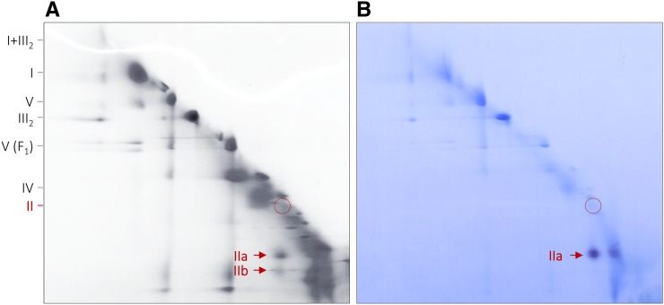
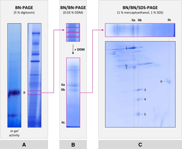
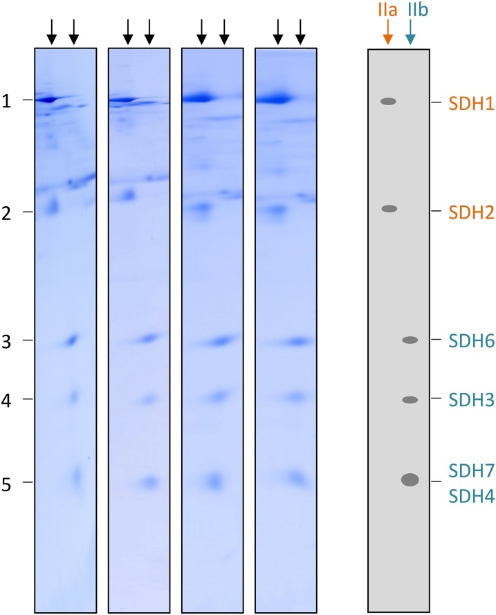
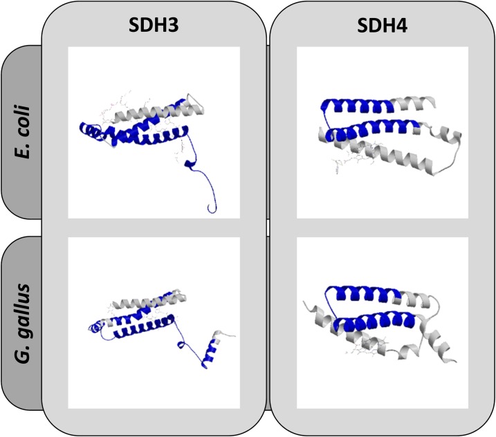
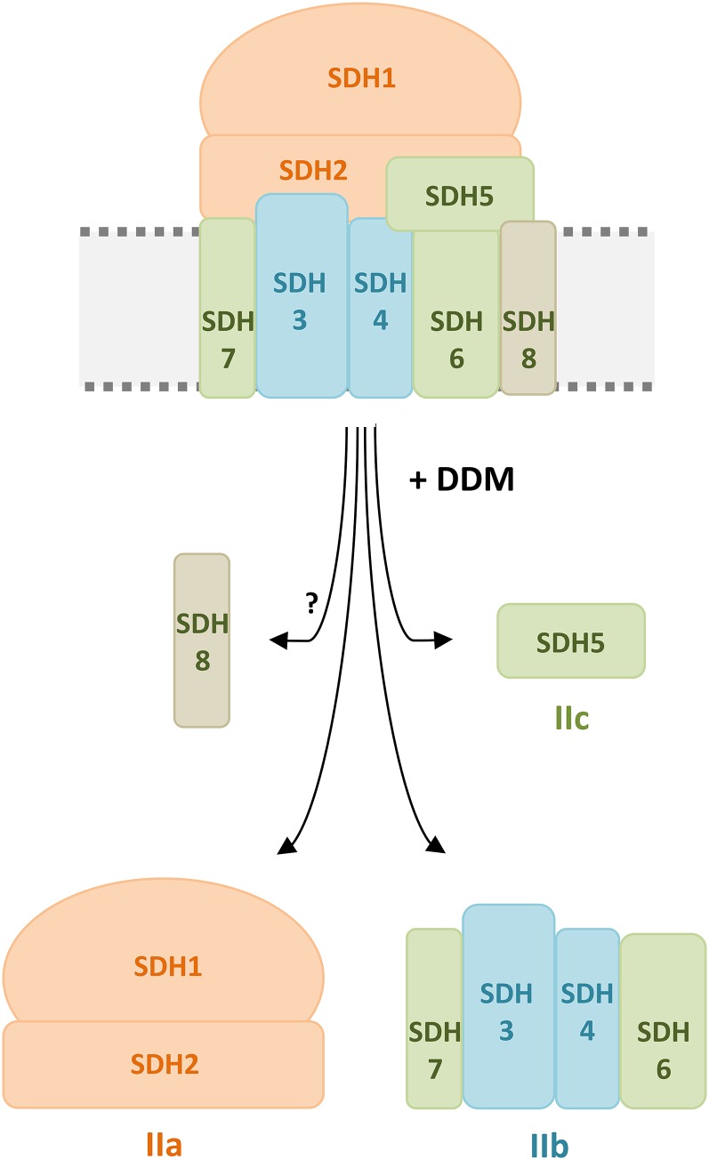
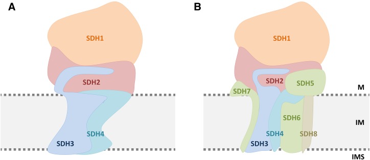
The succinate dehydrogenase complex (complex II) is a highly conserved protein complex composed of the SDH1 to SDH4 subunits in bacteria and in the mitochondria of animals and fungi. The reason for the occurrence of up to four additional subunits in complex II of plants, termed SDH5 to SDH8, so far is a mystery. Here, we present a biochemical approach to investigate the internal subunit arrangement of Arabidopsis (Arabidopsis thaliana) complex II. Using low-concentration detergent treatments, the holo complex is dissected into subcomplexes that are analyzed by a three-dimensional gel electrophoresis system. Protein identifications by mass spectrometry revealed that the largest subcomplex (IIa) represents the succinate dehydrogenase domain composed of SDH1 and SDH2. Another subcomplex (IIb) is composed of the SDH3, SDH4, SDH6, and SDH7 subunits. All four proteins include transmembrane helices and together form the membrane anchor of complex II. Sequence analysis revealed that SDH3 and SDH4 lack helices conserved in other organisms. Using homology modeling and phylogenetic analyses, we present evidence that SDH6 and SDH7 substitute missing sequence stretches of SDH3 and SDH4 in plants. Together with SDH5, which is liberated upon dissection of complex II into subcomplexes, SDH6 and SDH7 also add some hydrophilic mass to plant complex II, which possibly inserts further functions into this smallest protein complex of the oxidative phosphorylation system (which is not so small in plants).


Figures

Figure 1.
Figure 1.
Detection of complex II in mitochondrial fractions from Arabidopsis by BN/BN PAGE and complex II activity staining. BN/BN PAGE was carried out as outlined in “Materials and Methods.” A, Coomassie Blue-stained 2D gel. B, Parallel 2D gel used for complex II activity staining. The identities of the separated protein complexes and supercomplexes are given to the left: I, complex I; II, complex II; III2, dimeric complex III; IV, complex IV; V, complex V; V(F1), F1 part of complex V; I+III2, supercomplex formed by complexes I and III2. The position of intact complex II is given by the red circles, and the positions of two complex II subcomplexes (IIa and IIb) are indicated by red arrows.
Figure 2.
Figure 2.
Experimental strategy for analyzing complex II subcomplexes from Arabidopsis. A, Protein complexes of isolated mitochondria were separated by 1D BN PAGE and either complex II activity stained (left) or Coomassie Blue stained (right). The region of complex II as identified by activity staining (boxed in magenta) was cut out on parallel BN gel stripes that were neither fixed nor stained. B, Four BN gel pieces including native complex II were incubated with 0.03% (w/v) DDM and subsequently stacked on top of a second BN gel. This second BN PAGE dimension was used to separate complex II subcomplexes. C, The resulting gel stripe (boxed in magenta) was incubated with 1% (w/v) SDS and 1% (v/v) 2-mercaptoethanol, and subunits of complex II subcomplexes were subsequently separated in the orthogonal direction on a third gel dimension, which was carried out in the presence of SDS. Magenta arrows indicate the work flow. The position of native complex II and the positions of subcomplexes IIa, IIb, and IIc are indicated to the left or above the gel stripes. Proteins analyzed by mass spectrometry are numbered (for results, see Table I).
Figure 3.
Figure 3.
Subunit composition of complex II subcomplexes IIa and IIb in Arabidopsis. Subcomplexes of complex II were analyzed by BN/BN SDS-PAGE in four replicates. Subunits of subcomplexes IIa (orange) and IIb (turquoise) were identified by mass spectrometry (Table I). Identities of the complex II subunits are given to the right.
Figure 4.
Figure 4.
Structural modeling of SDH3 and SDH4 from Arabidopsis. 3D structure models were deduced from protein homology modeling using reference structures of SDH3 and SDH4 from E. coli (RCSB 2ACZ) and chicken (RCSB 2H88). The predicted Arabidopsis structures (given in blue) and the reference structures (given in gray) are superimposed. The analysis was carried out with SWISS-MODEL.
Figure 5.
Figure 5.
Stoichiometry of complex II subunits in Arabidopsis. Isolated mitochondria from Arabidopsis were solubilized by 5% digitonin, and mitochondrial proteins were separated by 2D BN SDS-PAGE. Stripes of the 2D gels including complex II (left, large subunits; right, small subunits) were stained using colloidal Coomassie Blue (left), Krypton fluorescent protein stain (middle), or SYPRO Ruby protein stain (right). Gel stripes were scanned using a Typhoon laser scanner, and proteins were quantified by densitometric measurements using the ImageJ software tool. The peak area-to-molecular mass ratio was calculated for SDH2, SDH5, SDH6, and SDH3 as well as for the protein spot including SDH4 and SDH7 (these two subunits overlap on the gel stripes). Results were normalized with respect to SDH3. Resulting stoichiometries are given at the bottom (highlighted in blue). Subunits not belonging to complex II are marked by asterisks.
Figure 6.
Figure 6.
Comparative analysis of OXPHOS complexes from Polytomella spp. and Arabidopsis. Isolated mitochondria from Polytomella spp. (A) and Arabidopsis suspension cell culture (B) were solubilized by 5% digitonin, and resulting protein fractions were separated by 2D BN SDS-PAGE. Gels were stained using colloidal Coomassie Blue. The identity of OXPHOS complexes is given above the gels. SDH subunits are marked by arrowheads. Complex II subunits from Polytomella spp. were identified by mass spectrometry (Table IV). Complex II subunits from Arabidopsis were identified previously (Klodmann et al., 2011); protein spots represent (from top to bottom) SDH1, SDH2, SDH5, SDH6, SDH3, and SDH7/SDH4 (SDH8 is not visible but most likely is present in the dye front at the position indicated by the gray arrowhead in B). Identities of OXPHOS complexes are given above the 2D gels: I, complex I; I*, subcomplex of complex I; II, complex II; III2, dimeric complex III; IV, complex IV; V, monomeric complex V; V2, dimeric complex V; V2*, subcomplex of dimeric complex V; I+III2, supercomplex composed of complex I and dimeric complex III.
Figure 7.
Figure 7.
Controlled disassembly of complex II from Arabidopsis by detergent treatment. The scheme shows complex II disassembly induced by incubation with 0.03% (w/v) DDM. Upon detergent treatment, the complex is split into three subcomplexes termed IIa, IIb, and IIc. The largest subcomplex (IIa; orange) consists of the matrix-exposed subunits SDH1 and SDH2. A smaller subcomplex (IIb; turquoise) represents the membrane anchor of complex II. It consists of SDH3 and SDH4 as well as the plant-specific subunits SDH6 and SDH7 (green). The smallest subcomplex (IIc; green) consists of the plant-specific and hydrophilic subunit SDH5, which forms neither part of subcomplex IIa nor IIb. The localization of the plant-specific subunit SDH8 (beige) is not known. Note that not all subunit interactions shown in the scheme have been experimentally proven. SDH7 is suggested to interact with SDH3, and SDH6 is suggested to interact with SDH4, based on evolutionary considerations (Table III). Furthermore, SDH5 occurred during evolution in parallel with SDH6 and possibly is involved in its integration into complex II. However, SDH5 also could be positioned at another location.
Figure 8.
Figure 8.
Subunit arrangement within mitochondrial complex II of chicken (A) and Arabidopsis (B). The subunit model for chicken is taken from the crystal structure (Huang et al., 2006); the model for Arabidopsis is based on the biochemical results presented here (Figs. 2 and 3) as well as on amino acid sequence analyses (Fig. 4; Supplemental Figs. S2 and S5). The top part of the complex, which protrudes into the mitochondrial matrix (M), includes SDH1 (orange) and SDH2 (red). This domain contains the succinate-binding site and includes covalently bound FAD in SDH1 as well as three iron-sulfur clusters in SDH2. The bottom part of the complex anchors SDH1 and SDH2 to the inner mitochondrial membrane (IM). Besides SDH3 and SDH4 (blue), it includes the plant-specific subunits SDH6 and SDH7 (green). SDH7 is suggested to interact with SDH3, and SDH6 is suggested to interact with SDH4, based on evolutionary considerations (Table III). The localization of SDH5 (green), which also is a plant-specific component, is probably at the interface between the succinate dehydrogenase and the membrane domain but also could be at another position. The localization of SDH8 (beige), which is only 4.9 kD in size and includes one membrane-spanning helix, is not known. IMS, Mitochondrial intermembrane space.

References

    1. Adams KL, Palmer JD (2003) Evolution of mitochondrial gene content: gene loss and transfer to the nucleus. Mol Phylogenet Evol 29: 380–395 - PubMed
    1. Adams KL, Rosenblueth M, Qiu YL, Palmer JD (2001) Multiple losses and transfers to the nucleus of two mitochondrial succinate dehydrogenase genes during angiosperm evolution. Genetics 158: 1289–1300 - PMC - PubMed
    1. Adl SM, Simpson AGB, Lane CE, Lukes J, Bass D, Bowser SS, Brown MW, Burki F, Dunthorn M, Hampl V, et al. (2012) The revised classification of eukaryotes. J Eukaryot Microbiol 59: 429–493 - PMC - PubMed
    1. Araújo WL, Nunes-Nesi A, Osorio S, Usadel B, Fuentes D, Nagy R, Balbo I, Lehmann M, Studart-Witkowski C, Tohge T, et al. (2011) Antisense inhibition of the iron-sulphur subunit of succinate dehydrogenase enhances photosynthesis and growth in tomato via an organic acid-mediated effect on stomatal aperture. Plant Cell 23: 600–627 - PMC - PubMed
    1. Baerenfaller K, Grossmann J, Grobei MA, Hull R, Hirsch-Hoffmann M, Yalovsky S, Zimmermann P, Grossniklaus U, Gruissem W, Baginsky S (2008) Genome-scale proteomics reveals Arabidopsis thaliana gene models and proteome dynamics. Science 320: 938–941 - PubMed

Publication types

MeSH terms

LinkOut - more resources