. 2017 Mar 17;45(5):2704-2713.
doi: 10.1093/nar/gkw1293.

Topoisomerase 3β is the major topoisomerase for mRNAs and linked to neurodevelopment and mental dysfunction

Affiliations

Topoisomerase 3β is the major topoisomerase for mRNAs and linked to neurodevelopment and mental dysfunction

Muzammil Ahmad et al. Nucleic Acids Res. .

Abstract

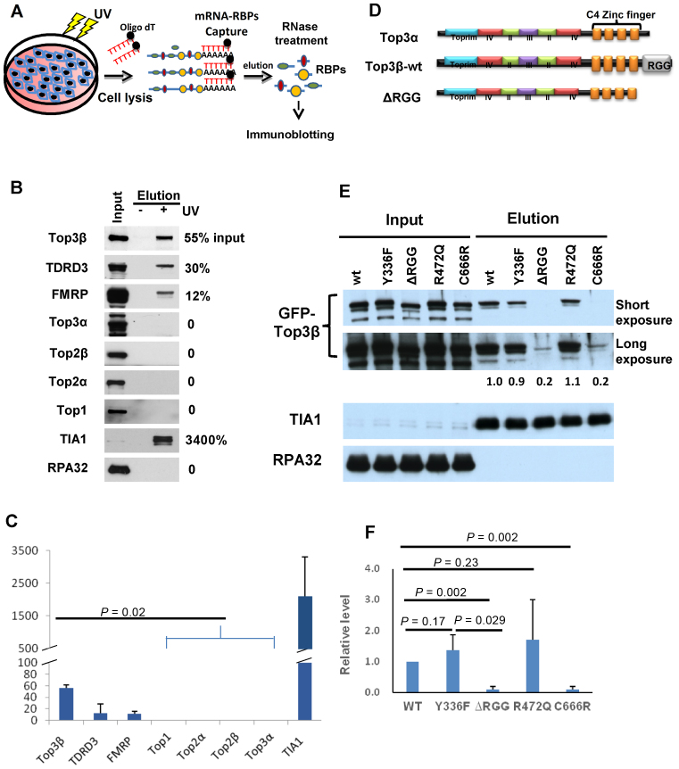
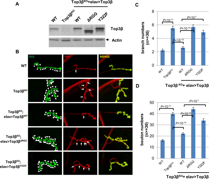
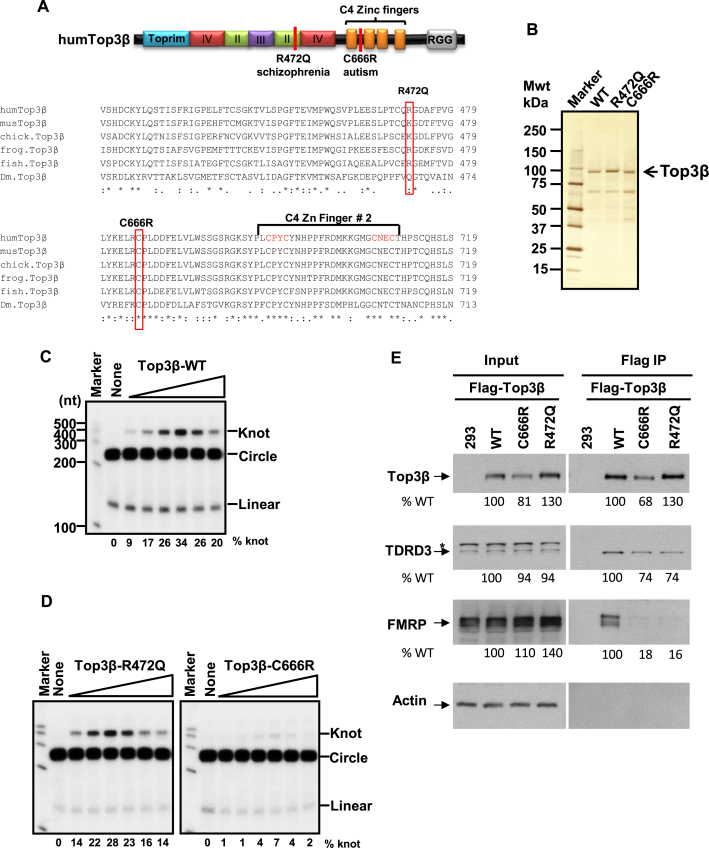
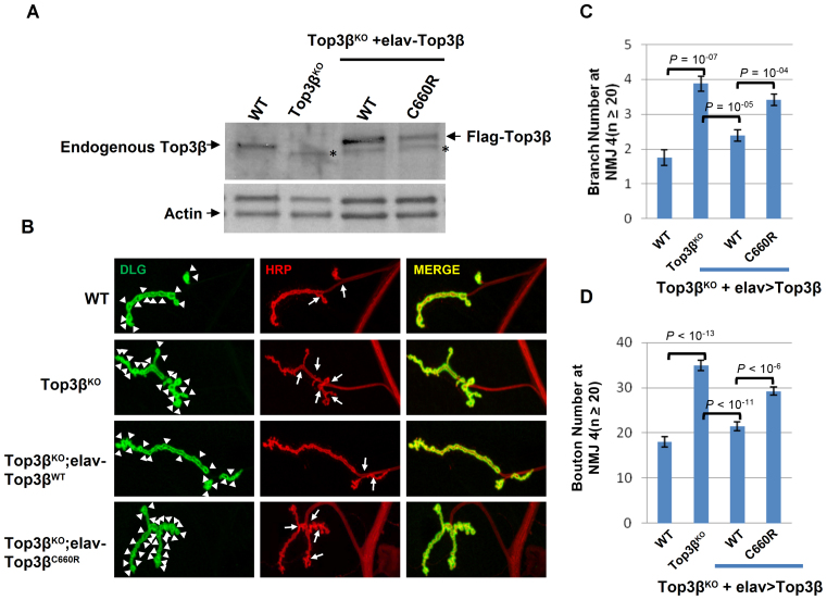
Human cells contain five topoisomerases in the nucleus and cytoplasm, but which one is the major topoisomerase for mRNAs is unclear. To date, Top3β is the only known topoisomerase that possesses RNA topoisomerase activity, binds mRNA translation machinery and interacts with an RNA-binding protein, FMRP, to promote synapse formation; and Top3β gene deletion has been linked to schizophrenia. Here, we show that Top3β is also the most abundant mRNA-binding topoisomerase in cells. Top3β, but not other topoisomerases, contains a distinctive RNA-binding domain; and deletion of this domain diminishes the amount of Top3β that associates with mRNAs, indicating that Top3β is specifically targeted to mRNAs by its RNA binding domain. Moreover, Top3β mutants lacking either its RNA-binding domain or catalytic residue fail to promote synapse formation, suggesting that Top3β requires both its mRNA-binding and catalytic activity to facilitate neurodevelopment. Notably, Top3β proteins bearing point mutations from schizophrenia and autism individuals are defective in association with FMRP; whereas one of the mutants is also deficient in binding mRNAs, catalyzing RNA topoisomerase reaction, and promoting synapse formation. Our data suggest that Top3β is the major topoisomerase for mRNAs, and requires both RNA binding and catalytic activity to promote neurodevelopment and prevent mental dysfunction.


Figures

Figure 1.
Figure 1.
Top3β is a major mRNA-binding topoisomerase and requires its RGG RNA-binding domain to bind mRNAs in vivo. (A) Schematic representation of an mRNA-binding protein capture assay modified from previous studies (21,22). mRNA and its RNA binding proteins (RBPs) were crosslinked by UV treatment in cells. After cell lysis, the mRNA-RBP complexes were captured by oligo-dT beads. After washing, the complexes were eluted and treated with RNase. The topoisomerases in RBPs were identified by immunoblotting. (B) Immunoblotting images and (C) quantification of mRNA-binding proteins captured on oligo-dT beads from lysates of HEK293 cells untreated or treated with UV. In (B), the relative ratios between the immunoblotting signals in the captured complex (lane 3) and the input (lane 1) were shown on the right. Only 0.1% of the input extract was loaded on the gel, whereas 50% of the eluted mRNA-bound RBPs were loaded. In (C), the graph shows the means of the relative ratios between the signals of the eluted proteins and the input from three independent experiments. The error bars indicate standard deviation. P-values between Top3β and other topoisomerases are calculated using Student's t-test. (D) Schematic representation of human Top3α, Top3β-wildtype and its RGG-deletion mutant (ΔRGG) proteins. The conserved core domains and the non-conserved CTDs, including Zn-fingers (orange boxes) and RGG-box, are indicated. Notably, the domain structure of Top3α and Top3β are highly similar, except the RGG-box which is only present in the former but not latter. (E) Immunoblotting images and (F) quantification from the mRNA-binding protein capture assay showing that GFP-tagged Top3β-ΔRGG and C666R mutants have strongly reduced mRNA binding activity after transfection in HEK293 cells, whereas Y336F and R472Q mutants retained the activity. In (B), for each Top3β mutant, the relative ratio of its immunoblotting signal in the captured mRNA complex versus the signal of the wild-type protein was shown between the images. A positive (TIA1) and negative (RPA) control was shown. In (F), the graph shows the means of the relative ratios between the immunoblotting signal of each mutant and that of the wild-type protein from three independent experiments. The error bars indicate standard deviation. P-values between Top3β and other topoisomerases are calculated using Student's t-test.
Figure 2.
Figure 2.
Top3β requires its distinctive RNA-binding domain and catalytic activity to promote synapse formation in Drosophila. (A) Immunoblotting shows the expression levels of Top3β wild type and various mutant proteins in brain extracts of Top3β-knockout and transgenic flies. (B) Representative immunofluorescence images of neuromuscular junctions at muscle 4 (NMJ4) of wandering third instar Drosophila larvae of different genotypes as indicated. The NMJ4 was co-labeled with a presynaptic marker (anti-HRP, red) and a postsynaptic marker (anti-DLG, green). The arrowheads mark synaptic boutons, and the arrows mark branches. Quantification of (C) synaptic branches and (D) boutons at NMJ4 from segments 3, 4 and 5 of both sides of wandering third instar larvae (n ≥ 30). The graphs show the means of the bouton or branch numbers, and error bars represent standard errors of mean from three independent experiments. The P-values shown above each bar were calculated using Student's t-test.
Figure 3.
Figure 3.
Two de novo single nucleotide variants (SNVs) of Top3β from schizophrenia and autism patients are defective in association with FMRP; and one SNV is also defective in RNA topoisomerase activity. (A) Schematic representation (top), and sequence alignment (bottom), of protein sequences of two de novo SNVs from schizophrenia and autism patients (17,18). Red colored vertical lines indicate locations of the mutations. Alignment of Top3β from several higher eukaryotic organisms was generated by Clustal W2.1. The identical residues are indicated by asterisks, whereas conserved residues are marked with columns. The species aligned are: human (gene identification (gi): 47678723), mouse (gi: 6755851), chicken (gi: 57525152), frog (gi: 148228452), fish (gi: 326667668), Drosophila (gi: 7290697). (B) A silver-stained gel shows purified recombinant human Top3β proteins of wild type and two de novo single nucleotide variants R472Q and C666R from a schizophrenia and autism patient, respectively. (C) Autoradiographs of the RNA topoisomerase assay of the wild type and the (D) two de novo SNVs of Top3β from schizophrenia (R472Q) and autism (C666R) patients. The closed circular substrate (circle), the knot product (knot) and linear RNA were indicated. The percentage of the knot converted from the circle was listed below each lane. These assays have been done three times, and the data are reproducible. (E) IP-western analyses show that Flag-Top3β-C666R and Flag-Top3β-R472Q co-immunoprecipitated with about 5-fold less amount of FMRP, and about 30% less amount of TDRD3 compared to the wild-type protein after transfection into HEK293 cells. Quantification of the immunoblotting images was performed using ImageJ software. The relative percentages of the input and IP signals for the two mutants were calculated using the signals of the wild-type protein as the standard (artificially set as 100%), and were listed below each image. The level of Flag-Top3β-C666R mutant is lower than that of the wild-type protein (about 20% less) in the input extract, suggesting that this mutation modestly reduces the stability of the protein. An asterisk marks a cross-reactive polypeptide. The experiments have been repeated twice, and the data are reproducible.
Figure 4.
Figure 4.
A de novo SNV of Top3β from an autism patient is defective in promoting synapse formation in Drosophila. (A) Western blot images show expression levels of Top3β wild type and mutant proteins in extracts of adult brains from the indicated genotypes. (B) Representative immunofluorescence images of NMJs at muscle 4 (NMJ4) of wandering third instar Drosophila larvae of different genotypes as indicated. The NMJ4 was co-labeled with a presynaptic marker (anti-HRP, red) and a postsynaptic marker (anti-DLG, green). The arrowheads mark synaptic boutons, and the arrows mark branches. (C and D) Quantification of synaptic branches and boutons at NMJ4 from segments 3, 4 and 5 of both sides of wandering third instar larvae (n ≥ 20). The graphs show the means of the boutons and branches, and error bars represent standard errors of mean from three independent experiments. The P-values shown above each bar were calculated using Student's t-test.

References

    1. Wang J.C. Cellular roles of DNA topoisomerases: a molecular perspective. Nat. Rev. Mol. Cell Biol. 2002; 3:430–440. - PubMed
    1. Pommier Y., Sun Y., Huang S.N., Nitiss J.L.. Roles of eukaryotic topoisomerases in transcription, replication and genomic stability. Nat. Rev. Mol. Cell Biol. 2016; 17:703–721. - PMC - PubMed
    1. Forterre P., Gribaldo S., Gadelle D., Serre M.C.. Origin and evolution of DNA topoisomerases. Biochimie. 2007; 89:427–446. - PubMed
    1. Stoll G., Pietilainen O.P., Linder B., Suvisaari J., Brosi C., Hennah W., Leppa V., Torniainen M., Ripatti S., Ala-Mello S. et al. Deletion of TOP3beta, a component of FMRP-containing mRNPs, contributes to neurodevelopmental disorders. Nat. Neurosci. 2013; 16:1228–1237. - PMC - PubMed
    1. Kwan K.Y., Greenwald R.J., Mohanty S., Sharpe A.H., Shaw A.C., Wang J.C.. Development of autoimmunity in mice lacking DNA topoisomerase 3beta. Proc. Natl. Acad. Sci. U.S.A. 2007; 104:9242–9247. - PMC - PubMed