. 2017 May;76(5):932-940.e3.
doi: 10.1016/j.jaad.2016.11.035. Epub 2016 Dec 29.

Human polyomavirus 6 and 7 are associated with pruritic and dyskeratotic dermatoses

Affiliations

Human polyomavirus 6 and 7 are associated with pruritic and dyskeratotic dermatoses

Khang D Nguyen et al. J Am Acad Dermatol. 2017 May.

Abstract

Background: Human polyomavirus (HPyV)6 and HPyV7 are shed chronically from human skin. HPyV7, but not HPyV6, has been linked to a pruritic skin eruption of immunosuppression.

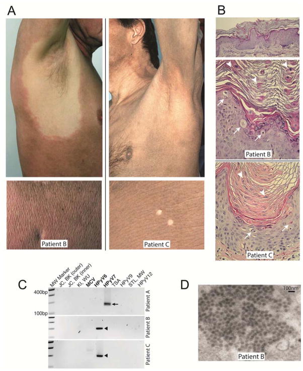
Objective: We determined whether biopsy specimens showing a characteristic pattern of dyskeratosis and parakeratosis might be associated with polyomavirus infection.

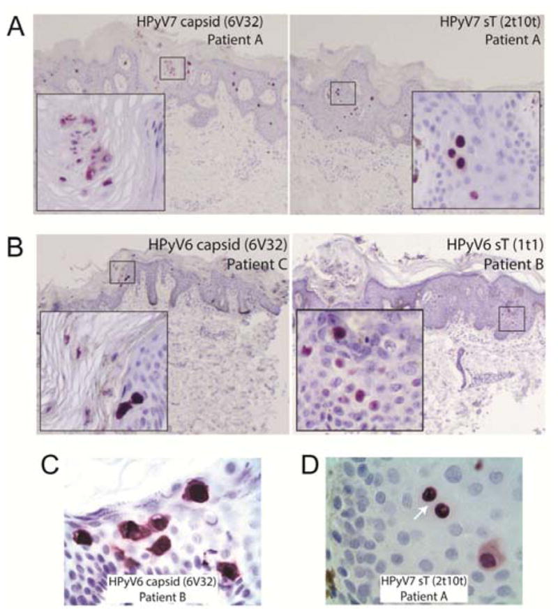
Methods: We screened biopsy specimens showing "peacock plumage" histology by polymerase chain reaction for HPyVs. Cases positive for HPyV6 or HPyV7 were then analyzed by immunohistochemistry, electron microscopy, immunofluorescence, quantitative polymerase chain reaction, and complete sequencing, including unbiased, next-generation sequencing.

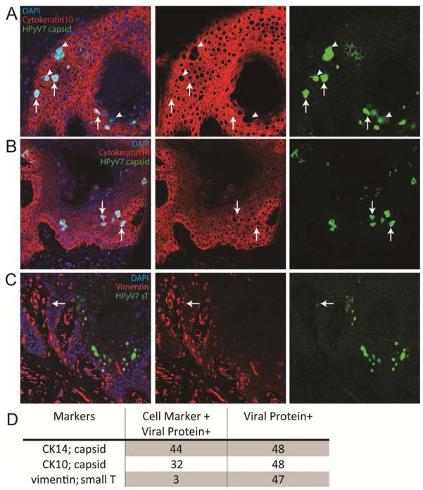
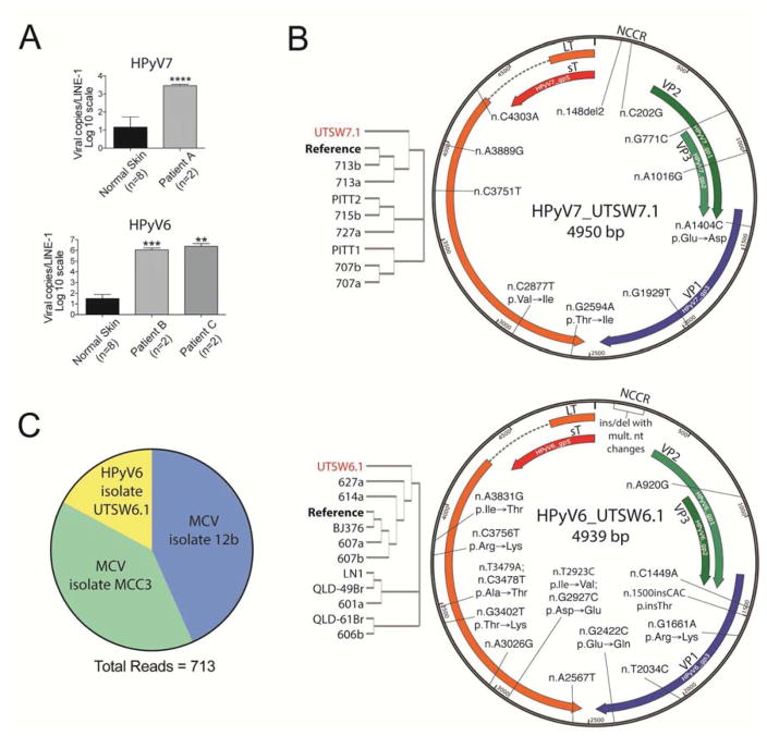
Results: We identified 3 additional cases of HPyV6 or HPyV7 skin infections. Expression of T antigen and viral capsid was abundant in lesional skin. Dual immunofluorescence staining experiments confirmed that HPyV7 primarily infects keratinocytes. High viral loads in lesional skin compared with normal-appearing skin and the identification of intact virions by both electron microscopy and next-generation sequencing support a role for active viral infections in these skin diseases.

Limitation: This was a small case series of archived materials.

Conclusion: We have found that HPyV6 and HPyV7 are associated with rare, pruritic skin eruptions with a distinctive histologic pattern and describe this entity as "HPyV6- and HPyV7-associated pruritic and dyskeratotic dermatoses."

Keywords: HIV/AIDS; dyskeratosis; human polyomavirus 6; human polyomavirus 7; immunosuppression; organ transplantation; parakeratosis; polyomavirus; pruritus.


Conflict of interest statement

Conflicts of interest: None declared.

Figures

Figure 1
Figure 1. HPyV6 and 7 are associated with a pruritic and dyskeratotic dermatosis
(A) Generalized, hyperpigmented, scaly eruptions in Patient B, with no known immunosuppression, and Patient C, a kidney/pancreas transplant patient, (Pock and Stork 2010). Images from an HIV/AIDS patient (Patient A) have previously been published (Champagne 2015); (B) From Patient B, on routine H&E sections, there is mild acanthosis and papillomatosis (top, H&E, 40X total magnification); several dyskeratotic cells are noted in the superficial epidermis (arrows), with eosinophilic, nucleated keratinocytes forming irregular columns of parakeratosis (arrowheads) in the stratum corneum (middle, H&E, 200X). From Patient C, an area with prominent columnar dyskeratosis showing dyskeratotic cells (arrows) and nucleated keratinocytes in the stratum corneum (arrowheads) (bottom, H&E, 200X) (C) PCR for human polyomaviruses yielded specific bands for HPyV7 (arrow) and HPyV6 (black arrowhead). (D) Electron microscopy of affected cells revealed numerous cytoplasmic ~40-nm icosahedral virions from Patient B (18000X, original direct magnification).
Figure 2
Figure 2. Immunohistochemistry against HPyV6 and 7 viral proteins
(A) Sample A shows capsid (6V32) expression in the nucleus and cytoplasm of affected cells. HPyV7 small T antigen (2t10t) is detected more strongly in the nuclei of affected cells. Samples B and C show capsid and HPyV6 small T antigen (1t1) in affected cells (insets, 200X). (B) High power images of the immunohistochemical stains show capsid protein expression in the nucleus and cytoplasm of the keratinocytes with viral cytopathic changes (left, oil immersion, 1000X). (C) Small T antigen is detectable in adjacent cells attached by desmosomes (right, arrow, oil immersion, 1000X) confirming their identity as keratinocytes.
Figure 3
Figure 3. HPyV7 infects keratinocytes
(A) Dual immunofluorescence (IF) staining for Cytokeratin 10 (CK10) and capsid (6V32) confirms that most capsid expressing cells also express CK10 (arrows, 400X). However, there are several cells in which the expression of CK10 is undetectable (arrowheads). (B) Dual IF staining for Cytokeratin 14 (CK10) and capsid (6V32) confirms that almost all capsid expressing cells also express CK14 (arrows, 400X). (C) Dual IF staining for Vimentin and small T antigen (2t10t) identifies rare T antigen expressing cells that also express vimentin (arrow, 400X). (D) Quantitation of dual IF staining images.
Figure 4
Figure 4. Novel strains of HPyV6 and 7 are abundant in lesional skin but can still be detected in asymptomatic patients
(A) Quantitative PCR comparing levels of HPyV6 and 7 detected in normal skin compared to lesional skin normalized to LINE-1 repeats. (n=independent biopsy; error bars=SD; t-test; **p≤0.01, ***p≤0.001, ****p≤0.0001). (B) Schematic of HPyV7 isolate from Patient A (HPyV7_UTSW7.1) and HPyV6 isolate from Patient B (HPyV6_UTSW6.1) with labels indicating the approximate position of nucleotide and AA changes. Cladograms (left) indicate the phylogenetic relationship of the current strains with previously sequenced strains. (C) Patient B possessed subclinical infections of HPyV6 isolate UTSW6.1 and MCV isolates after clinical symptoms had resolved in 2016. Pie chart indicates the proportion of reads (300bp or longer) corresponding to the indicated virus.

References

    1. Gardner SD, Field AM, Coleman DV, Hulme B. New human papovavirus (B.K) isolated from urine after renal transplantation. Lancet. 1971;1:1253–7. - PubMed
    1. Padgett BL, Walker DL, ZuRhein GM, Eckroade RJ, Dessel BH. Cultivation of papova-like virus from human brain with progressive multifocal leucoencephalopathy. Lancet. 1971;1:1257–60. - PubMed
    1. DeCaprio JA, Garcea RL. A cornucopia of human polyomaviruses. Nature reviews Microbiology. 2013;11:264–76. - PMC - PubMed
    1. Feng H, Shuda M, Chang Y, Moore PS. Clonal integration of a polyomavirus in human Merkel cell carcinoma. Science. 2008;319:1096–100. - PMC - PubMed
    1. van der Meijden E, Janssens RW, Lauber C, Bouwes Bavinck JN, Gorbalenya AE, Feltkamp MC. Discovery of a new human polyomavirus associated with trichodysplasia spinulosa in an immunocompromized patient. PLoS Pathog. 2010;6:e1001024. - PMC - PubMed

MeSH terms

LinkOut - more resources