. 2017 Mar:79:12-22.
doi: 10.1016/j.mcn.2016.12.006. Epub 2016 Dec 29.

Extracellular vesicles of the blood-brain barrier: Role in the HIV-1 associated amyloid beta pathology

Affiliations

Extracellular vesicles of the blood-brain barrier: Role in the HIV-1 associated amyloid beta pathology

Ibolya E András et al. Mol Cell Neurosci. 2017 Mar.

Abstract

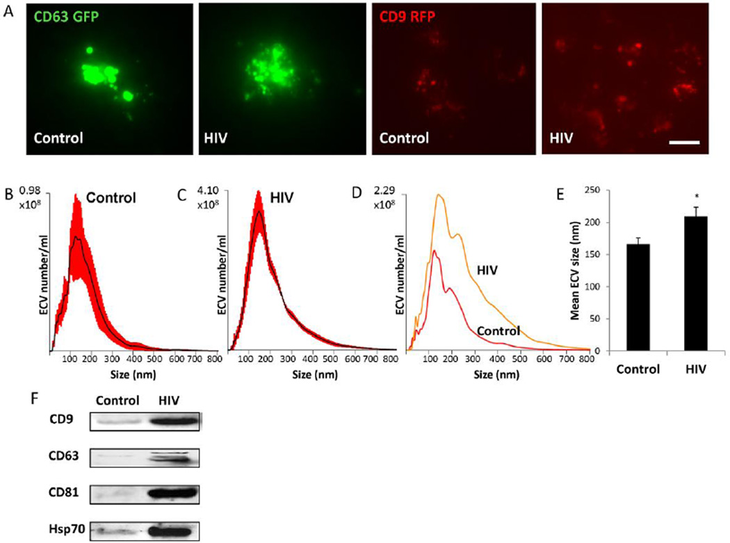
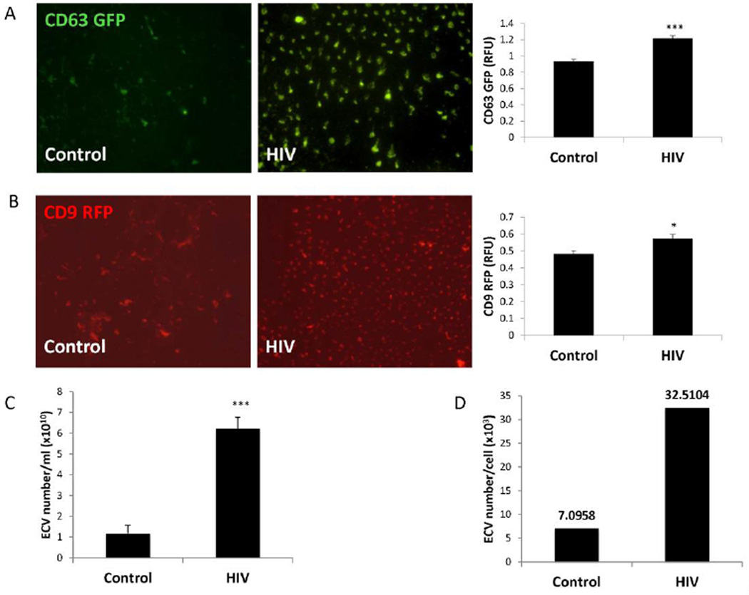
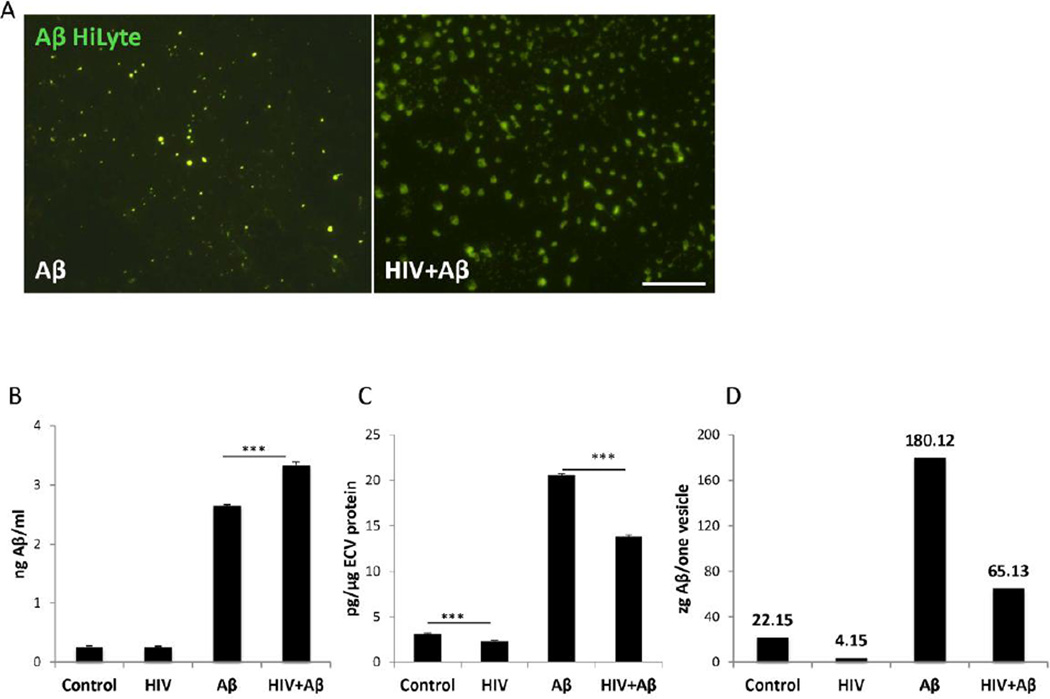
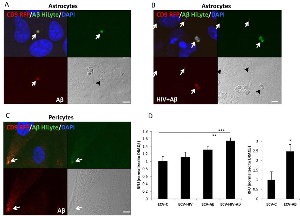
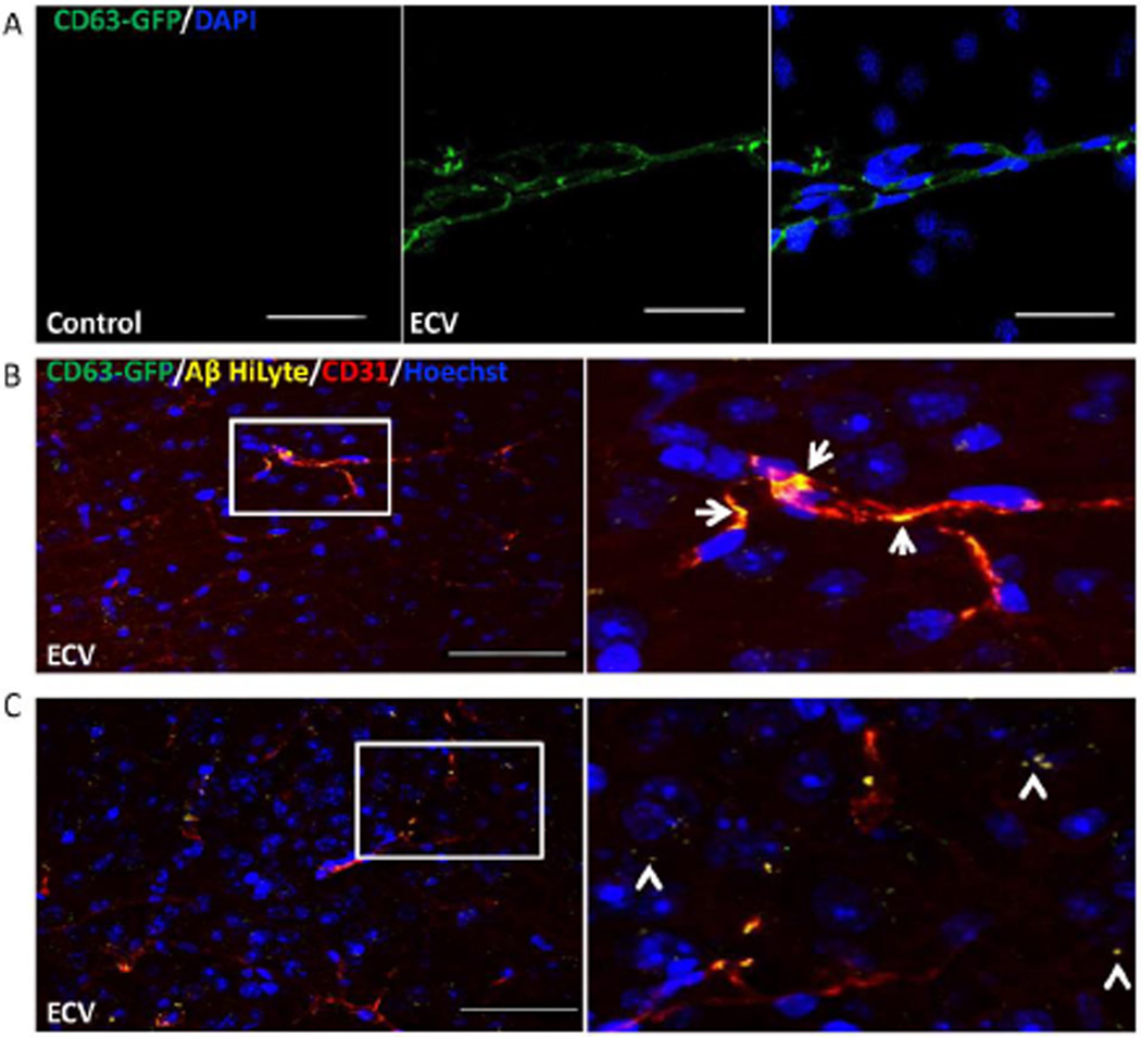
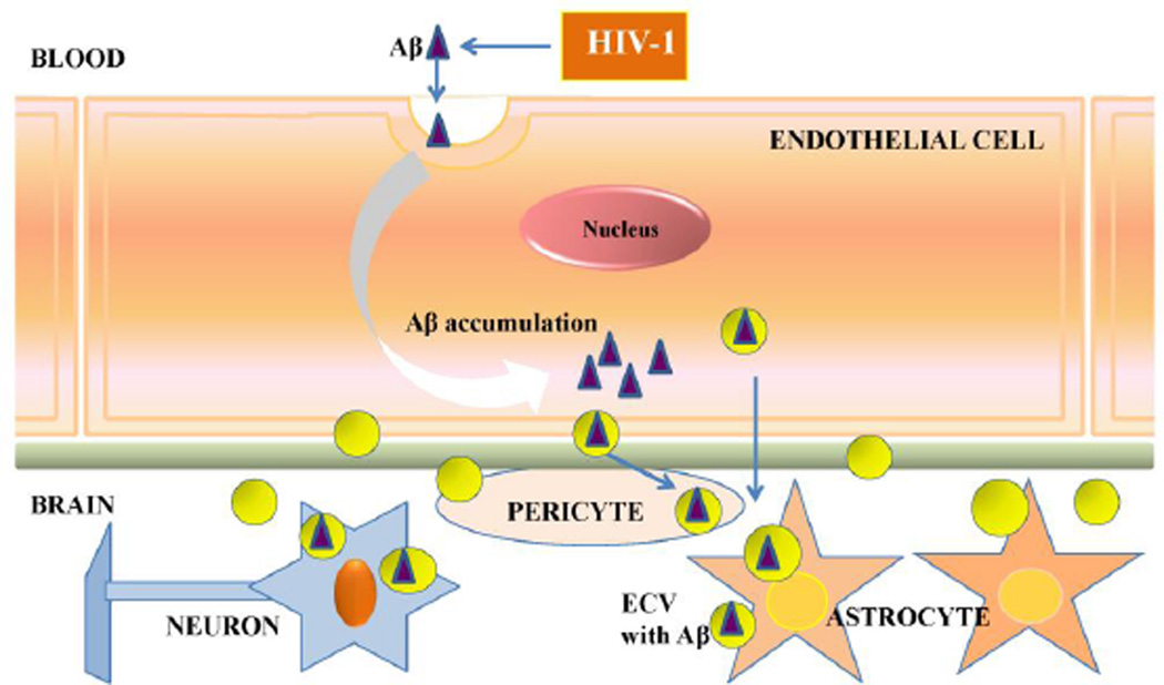
HIV-infected brains are characterized by increased amyloid beta (Aβ) deposition. It is believed that the blood-brain barrier (BBB) is critical for Aβ homeostasis and contributes to Aβ accumulation in the brain. Extracellular vesicles (ECV), like exosomes, recently gained a lot of attention as potentially playing a significant role in Aβ pathology. In addition, HIV-1 hijacks the exosomal pathway for budding and release. Therefore, we investigated the involvement of BBB-derived ECV in the HIV-1-induced Aβ pathology in the brain. Our results indicate that HIV-1 increases ECV release from brain endothelial cells as well as elevates their Aβ cargo when compared to controls. Interestingly, brain endothelial cell-derived ECV transferred Aβ to astrocytes and pericytes. Infusion of brain endothelial ECV carrying fluorescent Aβ into the internal carotid artery of mice resulted in Aβ fluorescence associated with brain microvessels and in the brain parenchyma. These results suggest that ECV carrying Aβ can be successfully transferred across the BBB into the brain. Based on these observations, we conclude that HIV-1 facilitates the shedding of brain endothelial ECV carrying Aβ; a process that may increase Aβ exposure of cells of neurovascular unit, and contribute to amyloid deposition in HIV-infected brain.

Keywords: Amyloid beta; Blood-brain barrier; Extracellular vesicles; HIV-1.


Figures

Figure 1
Figure 1. Characterization of extracellular vesicles (ECV) released from control and HIV-1 exposed brain endothelial cells
A) HBMEC were transfected with CD63 or CD9 Cyto-Tracer constructs (pT-CD63-GFP or pT-CD9-RFP), resulting in cells secreting green or red fluorescent ECV. The cells were exposed to 30 ng/ml HIV-1 particles for 48 h. The images represent live imaging of CD63-GFP or CD9-RFP positive ECV budding from control (left panels) and HIV-1 exposed cells (right panels). Scale bar: 20 µm. B and C) Size distribution of isolated ECV (±SEM are represented by red shading, n=4) from control and HIV-treated cultures, respectively. D) A comparison of representative graphs for the control and HIV groups illustrating the differences in size and amount. E) Quantitative analysis of mean ECV size in control and HIV-treated cultures (mean ± SEM, n=4). * Statistically significant as compared to control at p<0.05. F) Expression of marker proteins CD9, CD63, CD81, Hsp70 in HBMEC-derived ECV as determined by western blot.
Figure 2
Figure 2. HIV-1 exposure increases brain endothelial ECV secretion
HBMEC were transfected with pT-CD63-GFP (A) or pT-CD9-RFP (B) and exposed to 30 ng/ml HIV-1 for 48 h. Green or red fluorescent ECV were isolated from the culture media, imaged by fluorescence microscopy (scale bar: 20 µm) and quantified using a plate reader. HIV-1 exposure resulted in an increase in CD63-GFP and CD9-RFP positive fluorescence. Values are mean ± SEM, n=14–16. C–D) Non-transected HBMEC were exposed to 30 ng/ml HIV-1 for 48 h, followed by isolation of ECV from the culture media and quantification by NTA. The analyses indicate total ECV number (C) and ECV particle number produced by individual parent cells (D) in control and HIV-1 treated cultures. Values are mean ± SEM, n=4. * Statistically significant as compared to control at p<0.05. ***Statistically significant as compared to control at p<0.001.
Figure 3
Figure 3. The impact of HIV-1 on Aβ levels in HBMEC-derived ECV
A) Visualization of Aβ (1–40) HiLyte Alexa Fluor488 in ECV isolated from media of HBMEC exposed to HIV (30 ng/ml) and/or 100 nM Aβ (1–40) HiLyte for 48 h. Note different sizes and number of ECV in control and HIV-treated cultures. Aβ (1–40) HiLyte (green fluorescence) was assessed by fluorescence microscopy. Representative images from three experiments. Scale bar: 20 µm. B–C) Aβ (1–40) levels in isolated ECV from media of HBMEC treated as in (A) was measured by ELISA and normalized to cell culture media volume (B) or to ECV protein content (C). D) Aβ (1–40) levels per vesicle (1 zeptogram=10−21 g). Values are mean ± SEM, n=4. ***Statistically significant as compared to control or to Aβ (1–40) HiLyte treated group at p<0.001.
Figure 4
Figure 4. Transfer of Aβ (1–40) HiLyte cargo from donor HBMEC-derived ECV to recipient cells of the neurovascular unit
HBMEC transfected with pT-CD9-RFP were exposed to HIV (30 ng/ml) and/or 100 nM Aβ (1–40) HiLyte Alexa Fluor488 for 48 h, followed by the isolation of ECV from the cell culture media and treatment of astrocytes (A and B) or pericytes (C) for 24 h. All images are performed by confocal microscopy. DAPI staining (blue) visualizes nuclei. Colocalization of CD9-RFP (red fluorescence) and Aβ (1–40) HiLyte (green fluorescence) in astrocytes cultures exposed to ECV from (A) Aβ-treated HBMEC and (B) HIV-1 plus Aβ-treated HBMEC. (C) Colocalization of CD9-RFP (red fluorescence) and Aβ (1–40) HiLyte (green fluorescence) in pericyte cultures exposed to ECV from Aβ-treated HBMEC. Representative images from three experiments. Scale bar: 5 µm. (D) Quantification of Aβ (1–40) HiLyte fluorescence in recipient astrocytes (left graph) and pericytes (right graph). HBMEC were exposed and ECV isolated as in A and B. Astrocytes and pericytes grown on 96-well plates were exposed to fluorescent HBMEC-derived ECV for 24 h. Controls were exposed to non-fluorescent ECV from HBMEC. After washing with PBS, Aβ (1–40) HiLyte fluorescence was measured (abs/em 503/528 nm) in a plate reader. The values were normalized to DRAQ5 fluorescence. Values are mean ± SEM, n=14–16. *Statistically significant as compared to control at p<0.05. **Statistically significant as compared to control at p<0.01. ***Statistically significant as compared to control at p<0.001.
Figure 5
Figure 5. ECV transfer Aβ across the BBB into the brain parenchyma
HBMEC transfected with pT-CD63-GFP were exposed to 100 nM Aβ (1–40) HiLyte AlexaFluor647 for 48 h, followed by isolation of ECV from cell culture media. 2.5×10⁷ ECV were infused into the mouse brain via the internal carotid artery. Control mice were infused with saline. Analyses were performed 1 h post infusion by confocal microscopy; DAPI or Hoechst staining (blue) visualizes the nuclei. A) CD63-GFP positive ECV were associated with the isolated brain microvessels. Scale bar: 50 µm. B) Co-localization of CD63-GFP (green), Aβ (1–40) HiLyte AlexaFluor647 (yellow), and CD31 (endothelial marker, red) in the brain sections indicate association of CD63-GFP and Aβ with brain capillaries (arrows on the enlarged right panel). C) Brain sections were analyzed as in (B), indicating partial localization of Aβ in brain parenchyma and not associated with brain microvessels (arrowheads on the enlarged area), after an apparent crossing the BBB. Scale bar on B and C: 50 µm.
Figure 6
Figure 6. Schematic diagram of the HIV-1 induced Aβ exposure of the neurovascular unit via brain endothelial ECV
Our data indicate that HIV-1 facilitates the shedding of ECV from brain endothelial cells and increases ECV Aβ content. In addition, ECV can transfer Aβ to other cells of neurovascular unit, such as astrocytes and pericytes. These events may contribute to amyloid overload in HIV-infected brain, contributing to the development of neurocognitive dysfunction. Abbreviations: Aβ, amyloid beta; ECV, extracellular vesicles.

Similar articles

Cited by

References

    1. Abbott NJ. Astrocyte-endothelial interactions and blood-brain barrier permeability. J Anat. 2002;200(6):629–638. - PMC - PubMed
    1. Abbott NJ, Friedman A. Overview and introduction: the blood-brain barrier in health and disease. Epilepsia. 2012;53(Suppl 6):1–6. - PMC - PubMed
    1. Achim CL, et al. Increased accumulation of intraneuronal amyloid beta in HIV-infected patients. J Neuroimmune Pharmacol. 2009;4(2):190–199. - PMC - PubMed
    1. An K, et al. Exosomes neutralize synaptic-plasticity-disrupting activity of Abeta assemblies in vivo. Mol Brain. 2013;6:47. - PMC - PubMed
    1. Andras IE, et al. HIV-1-induced amyloid beta accumulation in brain endothelial cells is attenuated by simvastatin. Mol Cell Neurosci. 2010;43(2):232–243. - PMC - PubMed

Publication types

Substances