. 2017 Apr 1;19(4):514-523.
doi: 10.1093/neuonc/now232.

REST represses miR-124 and miR-203 to regulate distinct oncogenic properties of glioblastoma stem cells

Affiliations

REST represses miR-124 and miR-203 to regulate distinct oncogenic properties of glioblastoma stem cells

Anantha L Marisetty et al. Neuro Oncol. .

Abstract

Background: Glioblastoma (GBM) is one of the most common, aggressive, and invasive human brain tumors. There are few reliable mechanism-based therapeutic approaches for GBM patients. The transcriptional repressor RE1 silencing transcriptional factor (REST) regulates the oncogenic properties of a class of GBM stem-like cells (high-REST [HR]-GSCs) in humans. However, it has been unclear whether REST represses specific targets to regulate specific oncogenic functions or represses all targets with overlapping functions in GSCs.

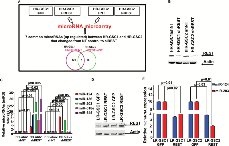
Methods: We used genome-wide, biochemical, and mouse intracranial tumorigenic assays to identify and determine functions of microRNA (miR) targets of REST in 2 independent HR-GSC lines.

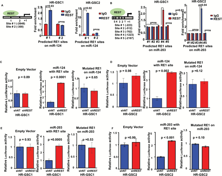
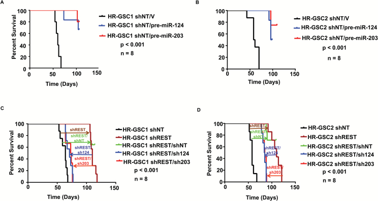
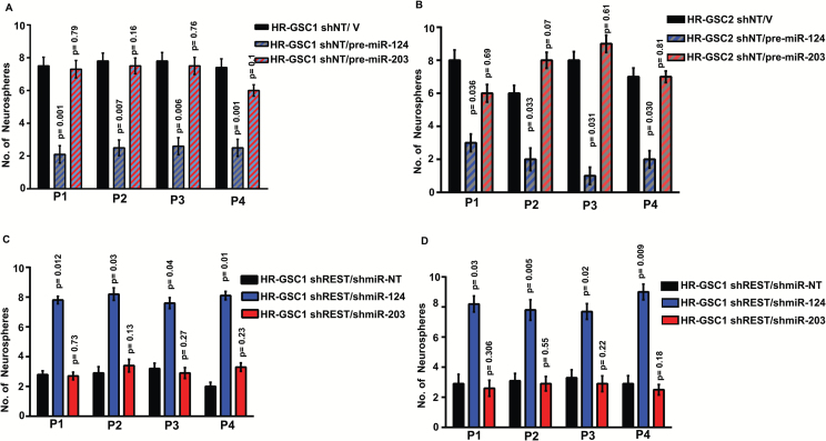
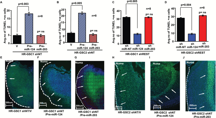
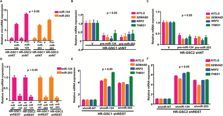
Results: Here we show that REST represses 2 major miR gene targets in HR-GSCs: miR-203, a new target, and miR-124, a known target. Gain of function of miR-124 or miR-203 in HR-GSCs increased survival in tumor-bearing mice. Importantly, the increased survival of tumor-bearing mice caused by knockdown of REST in HR-GSCs was reversed by double knockdown of REST and either miR-203 or miR-124, indicating that these 2 miRs are critical tumor suppressors that are repressed in REST-mediated tumorigenesis. We further show that while miR-124 and the REST-miR-124 pathways regulate self-renewal, apoptosis and invasion, miR-203 and the REST-miR-203 pathways regulate only invasion. We further identify and validate potential mRNA targets of miR-203 and miR-124 in REST-mediated HR-GSC tumor invasion.

Conclusions: These findings indicate that REST regulates its miR gene targets with overlapping functions and suggest how REST maintains oncogenic competence in GSCs. These mechanisms could potentially be utilized to block REST-mediated GBM tumorigenesis.

Keywords: GSCs; REST; apoptosis; invasion; miR-124; miR-203; survival.


Figures

Fig. 1
Fig. 1
Identification of miR-124 and miR-203 as REST targets in GSCs. (A, B) Genome-wide microRNA expression analysis. (A) Schematic representation of genome-wide microRNA expression analysis of the 2 HR-GSC cell lines representing REST loss-of-function using siREST manipulations (a total of 4 lines) resulted in only 7 microRNAs common between HR-GSC1 and HR-GSC2 in which REST expression differed between the nontargeting control (siNT) and siREST groups. (B–E) Functional validation of REST–miR-124 and REST–miR-203 pathways. (B, C) Loss-of-function manipulations: only 2 of the 7 microRNAs—miR-203 and miR-124—were significantly upregulated upon REST knockdown in both HR-GSC lines. (B) Stable cell lines were generated using lentiviruses carrying either the NT control or shREST and knockdown of REST protein was confirmed by western blotting assays. (C) MiR expression levels were determined using RT-qPCR. (D, E) Gain-of-function manipulations: expression levels of both miR-124 and miR-203 were decreased upon expression of exogenous REST. Stable lines of LR-GSC1 and LR-GSC2, which express low levels of the REST protein, were infected with retroviruses carrying either green fluorescent protein or exogenous REST gene and the expression levels of REST determined by western blotting assays (D). Analysis of the resulting cells by RT-qPCR showed that overexpression of REST causes lowered miR-124 and miR-203 expression in both the LR-GSC lines (E).
Fig. 2
Fig. 2
REST directly targets miR-124 and miR-203 in GSCs through specific sites present in their gene chromatin. (A, B) REST binds to miR-124 (A) and miR-203 (B) gene chromatins in both HR-GSC1 and HR-GSC2. Chromatin immunoprecipitation analysis was performed using either REST or immunoglobulin G (control) antibody followed by RT-qPCR using primers corresponding to potential RE1 sites present on the miR-124 gene chromatin (site #1 at -2648 upstream of the transcriptional start site and site #2, a random site, at -512) and miR-203 gene chromatin (site #1 at -223, site #2 at -512, site #3 at -762, and site #4 at -1223, and a random site #5 at -2167). REST binds to specific RE1 sites on these miR genes. (C–F) REST represses both miR-124 and miR-203 gene expression through the RE1 site present in its gene chromatin. We performed reporter gene analysis using a plasmid containing a luciferase gene downstream of either the specific REST binding site present on the miR-124 (C, D) or miR-203 (E, F) gene chromatins, a mutated version of the sites, or no site, and transfected the resulting plasmids into HR-GSC1/shNT and HR-GSC1/shREST (C, E), and HR-GSC2/shNT and HR-GSC2/shREST (D, F) cells and measured luciferase activity. Results showed that while the luciferase activity remained primarily unaltered when the plasmid with no site was expressed in either shNT- or shREST-expressing cells, it increased in shREST- compared with shNT-expressing cells when the plasmid contained the REST binding site. This increase in luciferase activity in shREST cells was reversed when the plasmid contained the mutated REST binding site.
Fig. 3
Fig. 3
MiR-124, miR-203, REST-miR-124, and REST-miR-203 pathways regulate survival of mice harboring brain tumors derived from both HR-GSC1 and HR-GSC2 cells. Kaplan–Meier survival plots of mice harboring HR-GSC1.shNT/V, HR-GSC1.shNT/pre-miR-124, and HR-GSC1.shNT/pre-miR-203 (A) and HR-GSC2.shNT/V, HR-GSC2.shNT/pre-miR-124, and HR-GSC2.shNT/pre-miR-203 (B) cells show that overexpression of either miR-124 or miR-203 in either HR-GSC1 or HR-GSC2 cells increased the survival of tumor-bearing mice, indicating that both miR-124 and miR-203 have tumor-suppressor functions in GSCs. Kaplan–Meier survival plots of mice harboring HR-GSC1 lines (HR-GSC1.shNT, HR-GSC1.shREST, HR-GSC1.shREST/shNT, HR-GSC1.shREST/shmiR-124, HR-GSC1.shREST/shmiR-203) (C) and HR-GSC2 lines (HR-GSC2.shNT, HR-GSC2.shREST, HR-GSC2.shREST/shNT, HR-GSC2.shREST/shmiR-124, HR-GSC2.shREST/shmiR-203) (D). Knockdown of REST by shREST in HR-GSC1 and HR-GSC2 cells caused increased survival compared with their shNT controls, as expected. Additional expression of shNT in these shREST-expressing cells did not significantly alter survival. In contrast, the double-knockdown shREST/shmiR-124 or shREST/shmiR-203 cells reversed the increased survival caused by single shREST in both the HR-GSC1 and HR-GSC2 cell lines, indicating that both the REST-miR-124 and REST-miR-203 pathways regulate the tumorigenesis of GSCs.
Fig. 4
Fig. 4
While MiR-124 and the RESTmiR-124 pathway regulate self-renewal of HR-GSCs, miR-203 and the RESTmiR-203 pathway do not. MiR-124 and miR-203 gain-of-function in HR-GSC1 cells (A: HR-GSC1.shNT/ pre-miR-124; HR-GSC1.shNT/ pre-miR-203) and HR-GSC2 cells (B: HR-GSC2.shNT/ pre-miR-124; HR-GSC2.shNT/ pre-miR-203) were subjected to neurosphere assays for 4 generations (P1–P4). While the exogenous miR-124 decreased self-renewal as compared with the control cells (HR-GSC1.shNT/Vector and HR-GSC2.shNT/Vector), the exogenous miR-203 did not. Knockdown of REST with shREST in either HR-GSC1 (C: HR-GSC1 shREST/shmiR-NT) or HR-GSC2 (D: HR-GSC2 shREST/shmiR-NT) caused lowered self-renewal, as was observed before (REF). Additional knockdown of miR-124 with shmiR-124 in either HR-GSC1 (C: HR-GSC1 shREST/shmiR-124) or HR-GSC2 (D: HR-GSC2 shREST/shmiR-NT) significantly rescued the lowered self-renewal cause by shREST alone in these cells. In contrast, shmiR-203 had negligible effect in double knockdown cells.
Fig. 5
Fig. 5
While miR-124 and the RESTmiR-124 pathway regulate both apoptosis and invasion, miR-203 and the RESTmiR-203 pathway regulate invasion in GSC-derived tumors in mouse brains. Quantification of double immunofluorescence staining of brain sections with anti-NuMA antibody that selectively stains human cells and TUNEL (A–D) showed increased apoptosis with the overexpression of miR-124 in HR-GSC1 (A) and HR-GSC2 (B) tumors and decreased apoptosis with knockdown of miR-124 in HR-GSC1/shREST (C) and HR-GSC2/shREST (D) tumors. In contrast, neither overexpression of miR-203 in either of the HR-GSC tumors (A and B, respectively) nor double knockdown of miR-203 in either the HR-GSC1/shREST (C) or HR-GSC2/shREST (D) tumors significantly altered tumor cell apoptosis. (E–J) Both MiR-124 and miR-203 regulate invasion in GSC-derived tumors in mouse brains. We stained the mouse brain tumor sections with anti-NuMA antibody and performed immunofluorescence analysis. Both HR-GSC1 and HR-GSC2 cells produced highly invasive tumors with cell invasion from the core of the tumor to the pial surface (E: HR-GSC1.shNT/Vector [V], H: HR-GSC2.shNT/Vector [V]), similar to what was seen previously. Overexpression of either miR-124 (F, I) or miR-203 (G, J) in both HR-GSC1 (F, G) and HR-GSC2 (I, J) tumors resulted in the blockade of invasion and the formation of circumscribed tumors (F: HR-GSC1.shNT/Pre-miR-124, G: HR-GSC1.shNT/Pre-miR-203, I: HR-GSC2.shNT/Pre-miR-124; J: HR-GSC2.shNT/Pre-miR-203).
Fig. 6
Fig. 6
Identification of downstream targets of RESTmicroRNA axis in GBM tumorigenesis. (A–C) Transcripts from mouse brain tumors generated from HR-GSC1/shNT (A, B) and HR-GSC2/shNT (A, C) cells expressing pre-miR-124, pre-miR-203, or the control vector were analyzed by RT-qPCR for the expression of miR-124 and miR-203 (A), KITLG, SEMA6D, NRP2, and THBS1 (B, C). (D–F) Transcripts from mouse brain tumors generated from HR-GSC1/shREST (D, E) and HR-GSC2/shREST (D, F) cells expressing either shmiR-124, shmiR-203, or shmiR-NT control were analyzed by RT-qPCR for the expression of miR-124 and miR-203 (D), KITLG, SEMA6D, NRP2, and THBS1 (D, F).

Similar articles

Cited by

References

    1. Aldape K, Zadeh G, Mansouri S, et al. Glioblastoma: pathology, molecular mechanisms and markers. Acta Neuropathol. 2015; 129(6):829–848. - PubMed
    1. Cloughesy TF, Cavenee WK, Mischel PS. Glioblastoma: from molecular pathology to targeted treatment. Annu Rev Pathol. 2014; 9:1–25. - PubMed
    1. Furnari FB, Cloughesy TF, Cavenee WK, et al. Heterogeneity of epidermal growth factor receptor signalling networks in glioblastoma. Nat Rev Cancer. 2015; 15(5):302–310. - PMC - PubMed
    1. Gladson CL, Prayson RA, Liu WM. The pathobiology of glioma tumors. Annu Rev Pathol. 2010; 5:33–50. - PMC - PubMed
    1. Wick W, Weller M, van den Bent M, et al. MGMT testing—the challenges for biomarker-based glioma treatment. Nat Rev Neurol. 2014; 10(7):372–385. - PubMed