. 2017 Jan 31;56(4):612-622.
doi: 10.1021/acs.biochem.6b00774. Epub 2017 Jan 17.

Quantification of the d-Ala-d-Lac-Terminated Peptidoglycan Structure in Vancomycin-Resistant Enterococcus faecalis Using a Combined Solid-State Nuclear Magnetic Resonance and Mass Spectrometry Analysis

Affiliations

Quantification of the d-Ala-d-Lac-Terminated Peptidoglycan Structure in Vancomycin-Resistant Enterococcus faecalis Using a Combined Solid-State Nuclear Magnetic Resonance and Mass Spectrometry Analysis

James D Chang et al. Biochemistry. .

Abstract

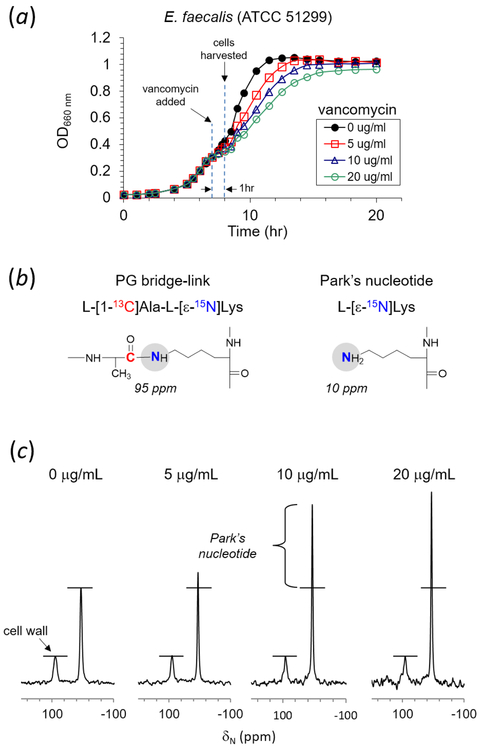
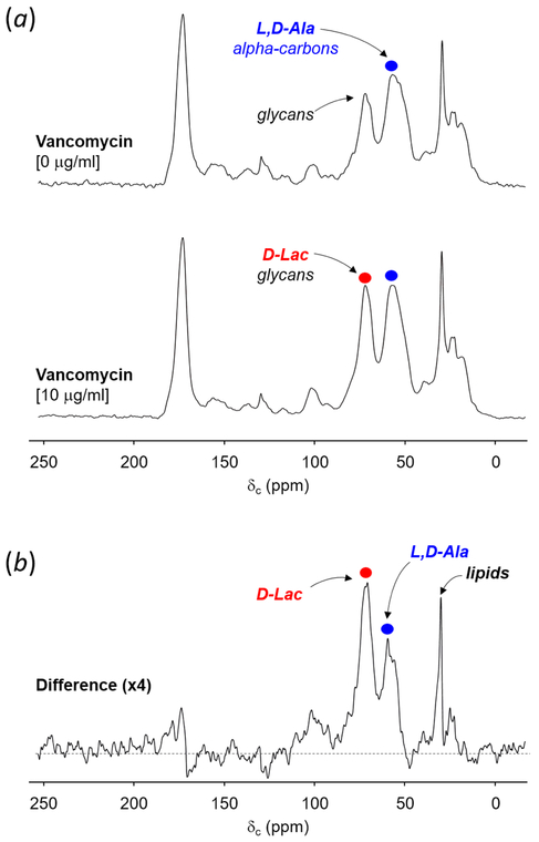
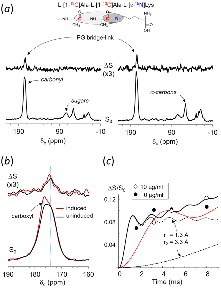
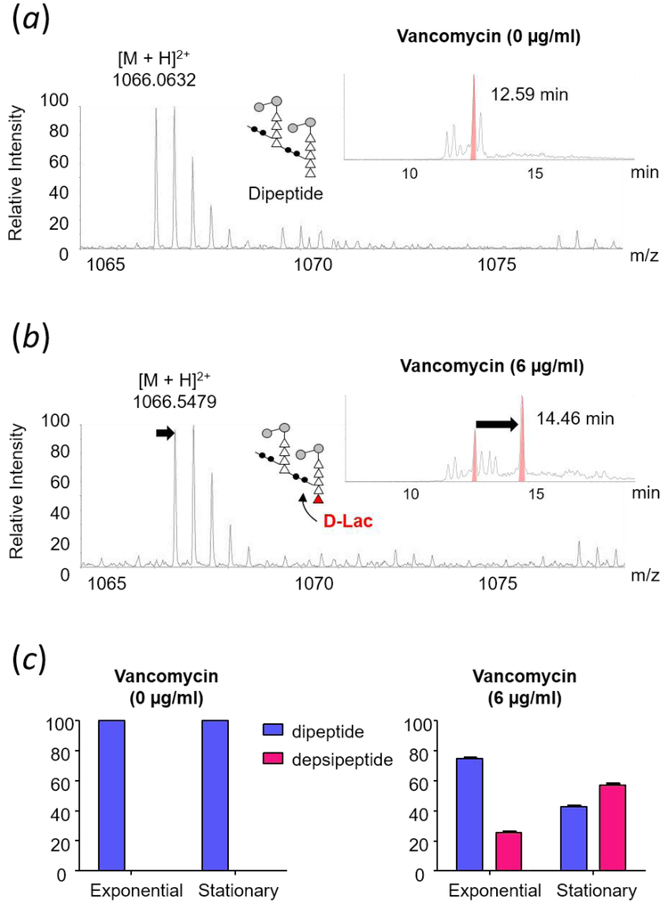
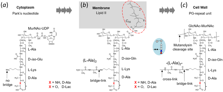
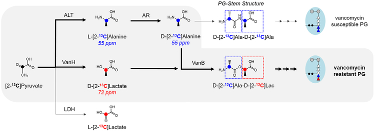
Induction of vancomycin resistance in vancomycin-resistant enterococci (VRE) involves replacement of the d-Ala-d-Ala terminus of peptidoglycan (PG) stems with d-Ala-d-Lac, dramatically reducing the binding affinity of vancomycin for lipid II. Effects from vancomycin resistance induction in Enterococcus faecalis (ATCC 51299) were characterized using a combined solid-state nuclear magnetic resonance (NMR) and liquid chromatography-mass spectrometry (LC-MS) analysis. Solid-state NMR directly measured the total amounts of d-Lac and l,d-Ala metabolized from [2-13C]pyruvate, accumulated Park's nucleotide, and changes to the PG bridge-linking density during the early exponential growth phase (OD660 = 0.4) in intact whole cells of VRE. A high level of accumulation of depsipeptide-substituted Park's nucleotide consistent with the inhibition of the transglycosylation step of PG biosynthesis during the initial phase of vancomycin resistance was observed, while no changes to the PG bridge-linking density following the induction of vancomycin resistance were detected. This indicated that the attachment of the PG bridge to lipid II by the peptidyl transferases was not inhibited by the d-Ala-d-Lac-substituted PG stem structure in VRE. Compositions of mutanolysin-digested isolated cell walls of VRE grown with and without vancomycin resistance induction were determined by LC-MS. Muropeptides with PG stems terminating in d-Ala-d-Lac were found only in VRE grown in the presence of vancomycin. Percentages of muropeptides with a pentapeptide stem terminating in d-Ala-d-Lac for VRE grown in the presence of vancomycin were 26% for the midexponential phase (OD660 = 0.6) and 57% for the stationary growth phase (OD660 = 1.0). These high percentages indicate that d-Ala-d-Lac-substituted lipid II was efficiently utilized for PG biosynthesis in VRE.


Figures

Figure 1.
Figure 1.
(a) Growth curves of vancomycin resistant E. faecalis (ATCC 51299) in ESM containing l-[1-13C]Ala and l-[ε-15N]Lys. Growth was monitored by measuring optical density at 660 nm (OD660nm). Vancomycin was added to final concentrations of 0, 5, 10, and 20 μg/mL during growth at OD660nm 0.3. Cells were harvested at OD660nm 0.4 for 15N-CPMAS NMR analysis. (b) Incorporation of l-[ε-15N]Lys into the PG bridge-link is visible as a lysyl-amide at 95 ppm, and into Park’s nucleotide as a lysyl-amine at 10 ppm in 15N-CPMAS spectra. (c) 15N-CPMAS spectra of E. faecalis labeled with l-[1-13C]Ala and l-[ε-15N]Lys. Spectra are normalized to 95-ppm intensity. l-[ε-15N]Lys incorporated into PG bridge-linked resonates at 95 ppm, and l-[ε-15N]Lys into proteins and Park’s nucleotide at 10 ppm. VRE grown in presence of vancomycin show Park’s nucleotide accumulation, indicating that vancomycin inhibited the transglycosylation step of PG biosynthesis. 15N-CPMAS spectra of VRE grown in vancomycin concentrations of 0, 5, 10, and 20 μg/mL are results of 74092, 80000, 70512, and 80000 accumulated scans, respectively. The magic angle spinning was at 5000 Hz. All measurements were carried out at ambient room temperature.
Figure 2.
Figure 2.
(a) 13C-CPMAS spectra of whole cells of E. faecalis grown in ESM supplemented with sodium-[2-13C]pyruvate (0.1 g/L). Vancomycin was added to final concentration of 10 μg/mL to one aliquot, and cells were grown for 1 hour and harvested. The 206-ppm peak assigned to [2-13C]pyruvate is absent from the 13C-CPMAS spectrum of VRE grown without vancomycin, due to [2-13C]pyruvate being fully utilized for the biosynthesis of [2-13C]Ala visible at 55 ppm (blue circle) and MurNAc at 72 ppm, which overlaps with the natural abundance 13C from glycans in PG. 13C-CPMAS spectra of VRE grown with and without vancomycin are results of 40000 and 80000 accumulated scans respectively. The magic angle spinning was at 5000 Hz. (b) The difference spectrum was obtained by subtracting 13C-CPMAS spectrum of VRE grown without vancomycin from the spectrum of VRE grown with vancomycin, and it shows increased d-[2-13C]Lac biosynthesis in VRE consistent with VanHB activity.
Figure 3.
Figure 3.
(a) 13C{15N}REDOR of whole cells of E. faecalis labeled with l-[1-13C]Ala and l-[ε-15N]Lys at 4.8 ms dipolar evolution grown in absence (left) and presence of vancomycin at 10 μg/mL (right). 13C{15N}REDOR spectra of VRE grown in vancomycin concentrations of 0 and 20 μg/mL are results of 75248 and 320000 accumulated scans respectively. The magic angle spinning was at 5000 Hz. (b) Enlarged overlaid S0 and ΔS spectra centered at 174 ppm for VRE grown with vancomycin (red), and without (black). ΔS 174-ppm peak intensity, which was directly proportional to the bridge-link density, was independent of induction of vancomycin resistance in VRE. (c) 13C{15N}REDOR dephasings (ΔS/S0) of alanyl-carboxyl carbon at 174 ppm. Solid line is the calculated REDOR dephasing curve for two 13Cs in l-[1-13C]Ala-l-[1-13C]Ala bridge that are dephased by the 15N of l-[ε-15N]Lys with 13C-15N distances of 1.3 and 3.3 Å (dotted lines). 13C{15N}REDOR dephasing is highly selective for the PG-bridge structure, as demonstrated by the calculated dephasing curve for 13C-15N distances corresponding to 2.0 and 4.0 Å (red curve), which does not fit observed dephasings.
Figure 4.
Figure 4.
Mass spectra of doubly-charged PG dimers identified from mutanolysin-digested isolated cell wall of vancomycin-resistant E. faecalis grown in absence (a) and presence of vancomycin (b) at stationary growth phase. The chemical structure, formula, and exact mass for PG dimers are provided in Supplementary Fig. S1. Select ion chromatograms (inset) show that D-Ala-D-Lac substituted PG dimer has a longer retention time that resolves it from the unmodified dimer. (c) Changes in the PG composition of dipeptide and depsipeptide terminated PG stems in the cell wall of E. faecalis grown in absence (left) and presence of vancomycin (right) during exponential and stationary growth phases. Muropeptides with PG stems terminating in d-Ala-d-Lac are only found in the cell wall of VRE grown in presence of vancomycin. Error bars represent 95% confidence interval.
Scheme 1.
Scheme 1.
Peptidoglycan biosynthesis in Enterococcus faecalis. (a) The first stage of PG biosynthesis takes place in the cytoplasm with Park’s nucleotide (UDP-MurNAc-l-Ala-D-iso-Glu-l-Lys-d-Ala-d-Ala) as the end product. In vancomycin-resistant E. faecalis, the terminal d-Ala of Park’s nucleotide is replaced by d-Lac. (b) The second stage of PG biosynthesis occurs on the interior side of bacterial cytoplasmic membrane, where UDP-MurNAc-stem from Park’s nucleotide is transported by the lipid transporter C55 to form lipid II (N-acetylglucosamine-N-acetyl-muramyl-pentapeptide-pyrophosphoryl-undecaprenol). Attachment of l-Ala-l-Ala bridge to the PG-stem is carried out by peptidyl transferases BppA1 and BppA2. d-iso-Glu is amidated to d-iso-Gln. (c) The final stage of PG biosynthesis is carried out on the external side of cytoplasmic membrane with incorporation of the PG-repeat unit into the cell wall.
Scheme 2.
Scheme 2.
[2-13C]Pyruvate metabolism in vancomycin-resistant E. faecalis. In absence of vancomycin, [2-13C]pyruvate is utilized for the biosynthesis of l-[2-13C]Ala by alanine transaminase (ALT), then to d-[2-13C]Ala by alanine racemase (AR). [2-13C]pyruvate metabolism to l-[2-13C]Lac by lactate dehydrogenase (LDH) does not occur in VRE under aerobic growth. Following the induction of vancomycin resistance, VanHB catalyzes the biosynthesis of d-[2-13C]Lac.

Similar articles

Cited by

References

    1. Arias CA, and Murray BE (2012) The rise of the Enterococcus: beyond vancomycin resistance, Nat. Rev. Microbiol 10, 266–278. - PMC - PubMed
    1. Cetinkaya Y, Falk P, and Mayhall CG (2000) Vancomycin-resistant enterococci, Clin. Microbiol. Rev 13, 686–707. - PMC - PubMed
    1. Singh M, Kim SJ, Sharif S, Preobrazhenskaya M, and Schaefer J (2015) REDOR constraints on the peptidoglycan lattice architecture of Staphylococcus aureus and its FemA mutant, Biochim. Biophys. Acta 1848, 363–368. - PMC - PubMed
    1. Kim SJ, Singh M, Sharif S, and Schaefer J (2014) Cross-link formation and peptidoglycan lattice assembly in the FemA mutant of Staphylococcus aureus, Biochemistry 53, 1420–1427. - PMC - PubMed
    1. Bouhss A, Josseaume N, Allanic D, Crouvoisier M, Gutmann L, Mainardi JL, Mengin-Lecreulx D, van Heijenoort J, and Arthur M (2001) Identification of the UDP-MurNAc-pentapeptide:L-alanine ligase for synthesis of branched peptidoglycan precursors in Enterococcus faecalis, J. Bacteriol 183, 5122–5127. - PMC - PubMed

Publication types

MeSH terms

LinkOut - more resources