

Bulbar conjunctival microvascular responses in dry eye
- PMID: 28042094
- PMCID: PMC5386830
- DOI: 10.1016/j.jtos.2016.12.002
Bulbar conjunctival microvascular responses in dry eye
Abstract
Purpose: Conjunctival microvascular responses may be a surrogate metric of efferent neural pathway function innervating the ocular surface as changes in blood flow occur within seconds after a stimulus. As somatosensory dysfunction may partially underlie dry eye (DE), in this study we evaluate whether bulbar conjunctival microvascular alterations correlate with various aspects of DE.
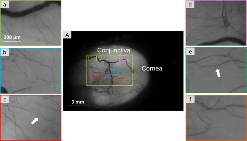
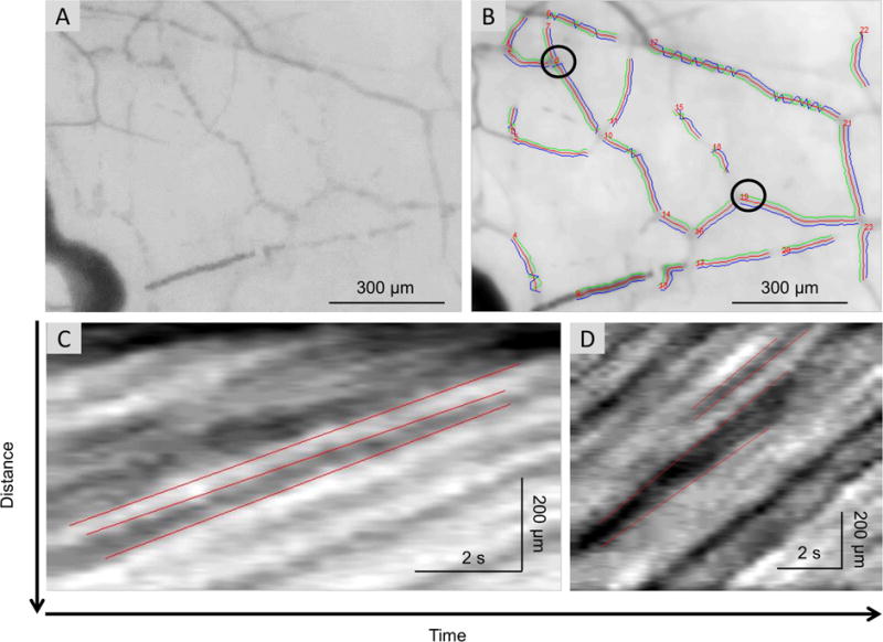
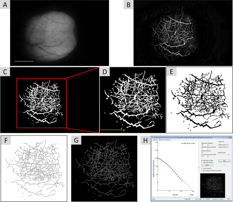
Methods: Fifty-six DE patients were prospectively recruited from a Veterans Affairs ophthalmology clinic over an 11-month period. DE symptoms and ocular pain were assessed along with DE signs. A novel functional slit lamp biomicroscope (FSLB) was used to image the temporal bulbar conjunctiva from the right eye before and after central corneal stimulation with an air puff. Blood flow velocities were measured and noninvasive microvascular perfusion maps (nMPMs) were created.
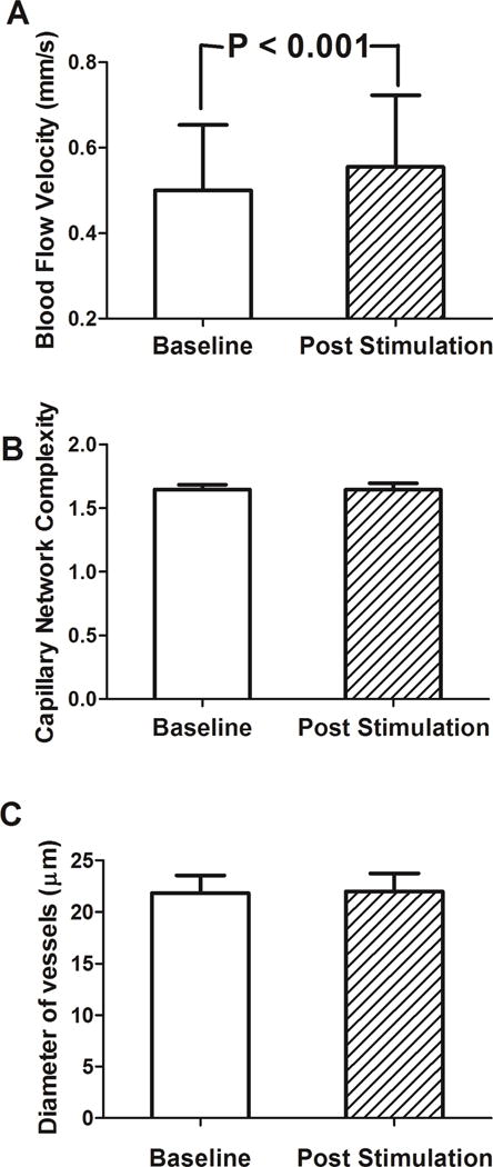
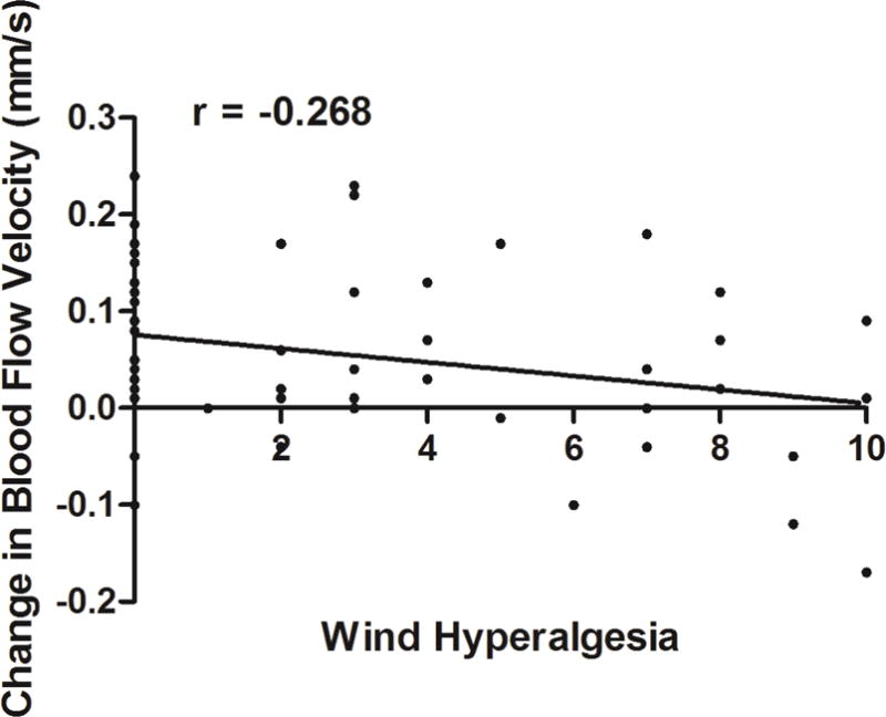
Results: The bulbar blood flow velocity was 0.50 ± 0.15 mm/s at baseline and increased to 0.55 ± 0.17 mm/s after stimulation (P < 0.001); the average change in velocity was 0.05 ± 0.09. nMPMs values and venule diameter, on the other hand, did not significantly increase after stimulation (1.64 ± 0.004 at baseline, 1.65 ± 0.04 after stimulation, P = 0.22 and 22.13 ± 1.84 μm at baseline, 22.21 ± 2.04 μm after stimulation, P = 0.73, respectively). Baseline blood flow velocity positively associated with Schirmer scores (r = 0.40, P = 0.002). Those with higher self-rated wind hyperalgesia demonstrated less change in blood flow velocity (r = -0.268, P = 0.046) after air stimulation on the central cornea.
Conclusion: Conjunctival blood flow velocity, but not vessel diameter or complexity, increases after wind stimuli. Baseline flow positively correlated with Schirmer scores while change in flow negatively correlated with self-reported wind hyperalgesia.
Keywords: Blood flow velocity; Corneal sensitivity; Fractal dimension; Functional slit lamp biomicroscopy; Neuropathic ocular pain.
Published by Elsevier Inc.
Conflict of interest statement
Figures
References
-
- Stern ME, Gao J, Siemasko KF, Beuerman RW, Pflugfelder SC. The role of the lacrimal functional unit in the pathophysiology of dry eye. Exp Eye Res. 2004;78:409–16. - PubMed
-
- The epidemiology of dry eye disease: report of the Epidemiology Subcommittee of the International Dry Eye WorkShop (2007) Ocul Surf. 2007;5:93–107. No authors listed. - PubMed
-
- Pouyeh B, Viteri E, Feuer W, Lee DJ, Florez H, Fabian JA, et al. Impact of ocular surface symptoms on quality of life in a United States veterans affairs population. Am J Ophthalmol. 2012;153:1061–66 e3. - PubMed
-
- Schein OD, Tielsch JM, Munoz B, Bandeen-Roche K, West S. Relation between signs and symptoms of dry eye in the elderly. A population-based perspective. Ophthalmology. 1997;104:1395–401. - PubMed
MeSH terms
Grants and funding
LinkOut - more resources
Full Text Sources
Other Literature Sources
Research Materials
