. 2017 Sep;31(9):1951-1961.
doi: 10.1038/leu.2016.393. Epub 2017 Feb 2.

Novel BET protein proteolysis-targeting chimera exerts superior lethal activity than bromodomain inhibitor (BETi) against post-myeloproliferative neoplasm secondary (s) AML cells

Affiliations

Novel BET protein proteolysis-targeting chimera exerts superior lethal activity than bromodomain inhibitor (BETi) against post-myeloproliferative neoplasm secondary (s) AML cells

D T Saenz et al. Leukemia. 2017 Sep.

Abstract

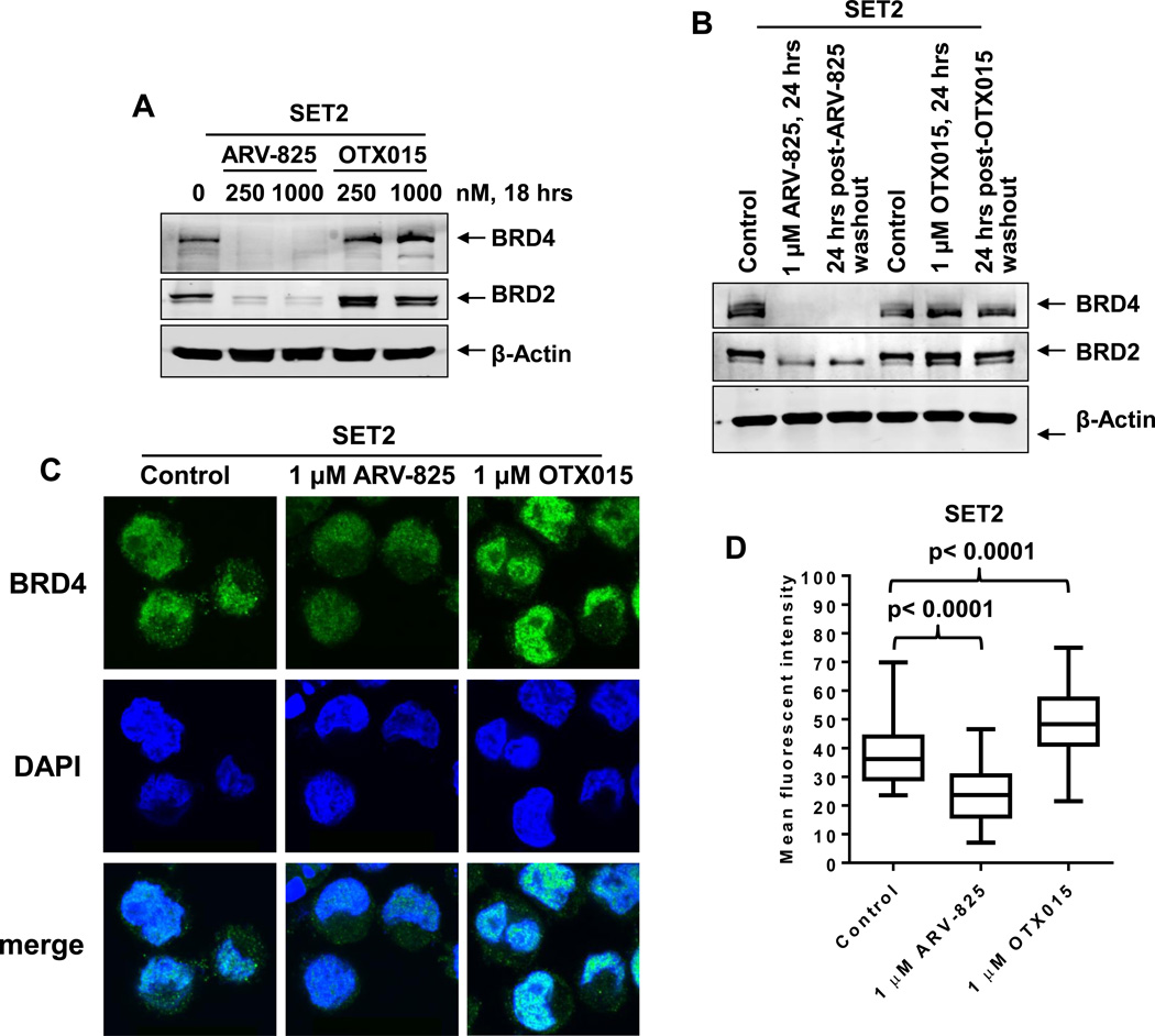
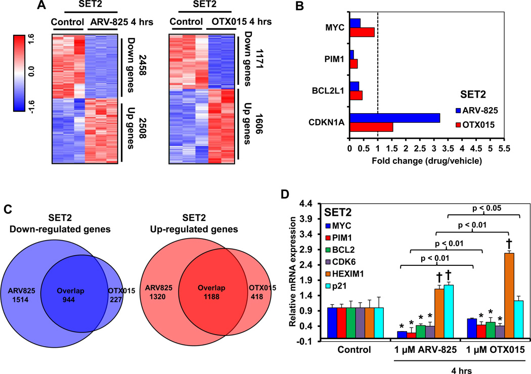
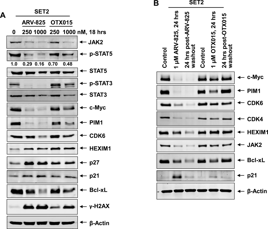
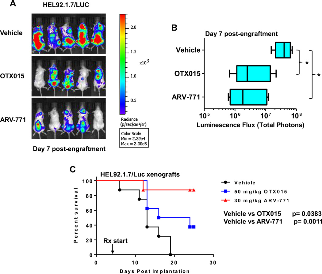
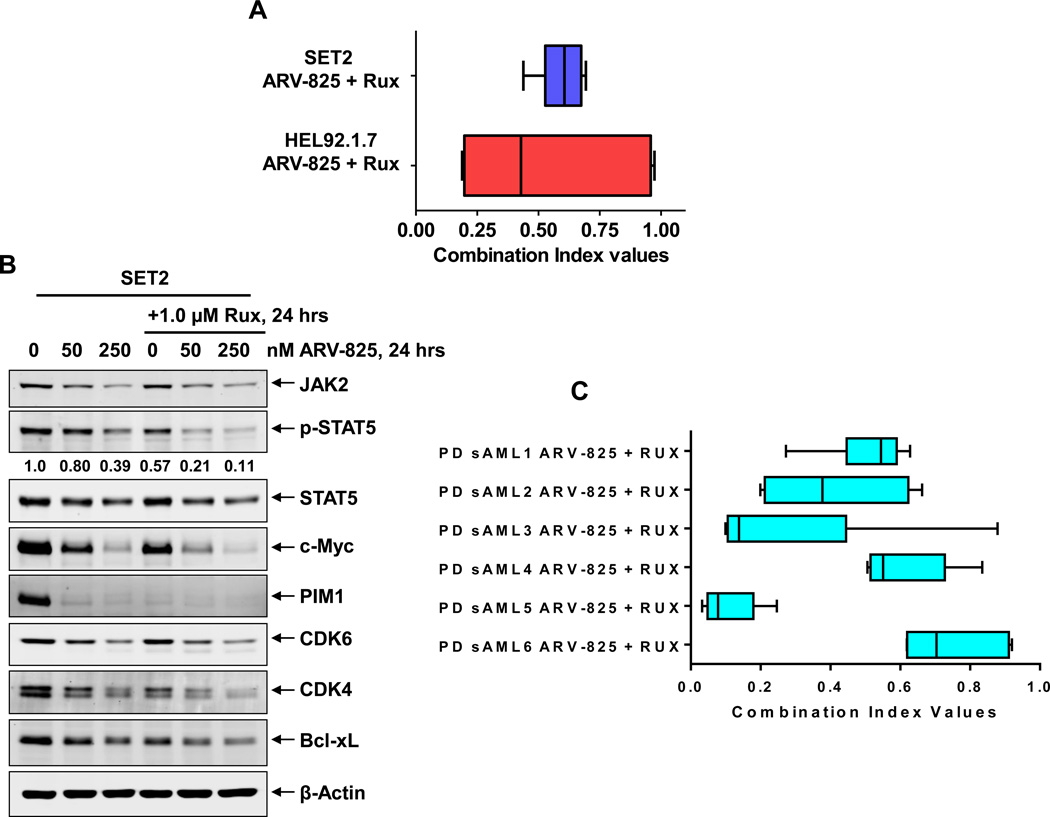
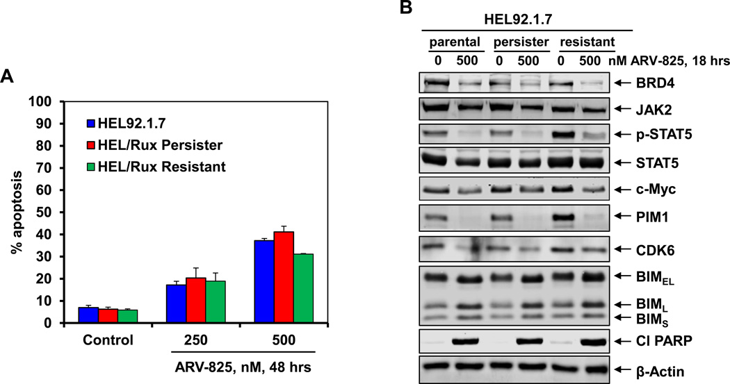
The PROTAC (proteolysis-targeting chimera) ARV-825 recruits bromodomain and extraterminal (BET) proteins to the E3 ubiquitin ligase cereblon, leading to degradation of BET proteins, including BRD4. Although the BET-protein inhibitor (BETi) OTX015 caused accumulation of BRD4, treatment with equimolar concentrations of ARV-825 caused sustained and profound depletion (>90%) of BRD4 and induced significantly more apoptosis in cultured and patient-derived (PD) CD34+ post-MPN sAML cells, while relatively sparing the CD34+ normal hematopoietic progenitor cells. RNA-Seq, Reverse Phase Protein Array and mass cytometry 'CyTOF' analyses demonstrated that ARV-825 caused greater perturbations in messenger RNA (mRNA) and protein expressions than OTX015 in sAML cells. Specifically, compared with OTX015, ARV-825 treatment caused more robust and sustained depletion of c-Myc, CDK4/6, JAK2, p-STAT3/5, PIM1 and Bcl-xL, while increasing the levels of p21 and p27. Compared with OTX015, PROTAC ARV-771 treatment caused greater reduction in leukemia burden and further improved survival of NSG mice engrafted with luciferase-expressing HEL92.1.7 cells. Co-treatment with ARV-825 and JAK inhibitor ruxolitinib was synergistically lethal against established and PD CD34+ sAML cells. Notably, ARV-825 induced high levels of apoptosis in the in vitro generated ruxolitinib-persister or ruxolitinib-resistant sAML cells. These findings strongly support the in vivo testing of the BRD4-PROTAC based combinations against post-MPN sAML.


Conflict of interest statement

Conflict of Interest: C.M.C. is the founder and Chief Scientific Advisor of, and possesses an equity ownership stake in, Arvinas, Inc. Y.Q., K.R. K.G.C., A.P.C., A.S. are Arvinas employees and possess an equity ownership stake in Arvinas. N.P. serves on the scientific advisory board of Incyte Pharmaceuticals. He has also served as a consultant and received research funding from Novartis Pharmaceuticals. All other authors state that they have no conflict of interest to declare.

Figures

Figure 1
Figure 1. Treatment with ARV-825 causes efficient and sustained depletion of BET protein expressions in sAML cells
A. SET-2 cells were treated with the indicated concentrations of ARV-825 or OTX015 for 18 hours. Total cell lysates were prepared and immunoblot analyses were conducted as indicated. The expression levels of β-Actin in the cell lysates served as the loading control. B. SET2 cells were treated with the indicated concentrations of ARV-825 or OTX015 for 24 hours. At the end of treatment, half of the cells were collected and snap frozen in liquid nitrogen. The remaining cells were washed three times in serum-free RPMI-1640 media to remove the drug (drug washout) and re-plated in complete media containing no drug for an additional 24 hours. Following this, total cell lysates were prepared and immunoblot analyses were conducted as indicated. The expression levels of β-Actin in the cell lysates served as the loading control. C. SET2 cells were treated with the indicated concentrations of ARV-825 or OTX015 for 16 hours. Then, cells were cytospun onto glass slides and immunostained for BRD4 expression. Cells were counterstained with DAPI and images were acquired utilizing confocal microscopy. Original magnification is 100X. D. Median fluorescent intensity of nuclear BRD4 staining in SET2 cells. At least 20 cells in each condition were quantified. Statistical differences in the median fluorescent intensity are noted on the boxplot.
Figure 2
Figure 2. Treatment with BET PROTAC, ARV-825 induces more apoptosis and loss of cell viability than BET protein inhibitor, OTX015 in cultured and patient-derived (PD) CD34+ sAML cells
A. SET-2 cells were treated with the indicated concentrations of ARV-825 or OTX015 for 48 hours. At the end of treatment, the % apoptotic cells were determined by flow cytometry. Columns, mean of three experiments; Bars, standard error of the mean. (The IC50 dose for ARV-825 is 14.5 ± 0.2 nM. The IC50 dose for OTX015 is 256 ± 21 nM. B. UKE1 cells were treated with the indicated concentrations of ARV-825 or OTX015 for 48 hours. At the end of treatment, the percentages of annexin V-positive, apoptotic cells were determined by flow cytometry. Columns, mean of three experiments; Bars, standard error of the mean. (The IC50 dose for ARV-825 is 256 ± 12.45 nM. The IC50 dose for OTX015 is > 500 nM. C. Patient-derived (PD) CD34+ sAML cells and normal CD34+ cells were treated with the indicated concentrations of ARV-825 or OTX015 for 48 hours. Following this, the % of propidium iodide-positive, non-viable cells was determined by flow cytometry. Columns, mean loss of cell viability from five PD samples; Bars, standard error of the mean.
Figure 3
Figure 3. Treatment with ARV-825 and OTX015 depletes the mRNA expression levels of c-Myc, PIM1, and BCL2L1/Bcl-xL but induces p21 protein expression levels in cultured and patient-derived sAML cells
A. SET2 cells were treated with 1 µM of ARV-825 or OTX015 for 4 hours. Samples shown represent biologic triplicates. RNA-Seq analysis was performed on the isolated RNA. The heat map shows the number of mRNAs with a fold-change exceeding 1.33X in either direction (relative to the untreated control), and p-value less than 0.05, following treatment with ARV-825 or OTX015. B. The graph shows the fold change (drug-treated divided by control) of selected gene targets in ARV-825 and OTX015-treated cells. Vertical line at 1.0 indicates no fold change. C. Venn diagram representing the expression signature overlap in down and upregulated genes cells following treatment with ARV-825 and OTX015 in SET2 cells. D. SET-2 cells were treated with the indicated concentrations of ARV-825 or OTX015 for 4 hours. Data shown represent biologic triplicates. At the end of treatment, total RNA was isolated and reverse transcribed. The resulting cDNA was used for real-time, quantitative PCR analysis. The relative mRNA expression of each target was normalized to GAPDH and compared to the untreated control cells. * indicates mRNA values that are significantly depleted compared to the untreated control cells (p <0.05). indicates mRNA values that are significantly induced compared to the untreated control cells (p <0.05).
Figure 4
Figure 4. Treatment with ARV-825 causes efficient and sustained depletion of BRD4 target gene expressions in sAML cells
A. SET-2 cells were treated with the indicated concentrations of ARV-825 or OTX015 for 18 hours. Total cell lysates were prepared and immunoblot analyses were conducted as indicated. The expression levels of β-Actin in the cell lysates served as the loading control. B. SET-2 cells were treated with the indicated concentrations of ARV-825 or OTX015 for 24 hours. At the end of treatment, half of the cells were collected, washed with 1X PBS and snap frozen in liquid nitrogen. The remaining cells were washed three times in serum-free RPMI-1640 media to remove the drug (drug washout) and re-plated in complete media containing no drug for an additional 24 hours. Following this, total cell lysates were prepared and immunoblot analyses were conducted as indicated. The expression levels of β-Actin in the cell lysates served as the loading control.
Figure 5
Figure 5. BET-PROTAC ARV-771 is more potent than BETi OTX015 in reducing the in vivo sAML burden and improving survival of NSG mice engrafted with luciferase transduced sAML cells
A. NSG mice were implanted with luciferase-expressing HEL92.1.7 cells and monitored for 4 days. Mice were imaged to document engraftment of leukemia and then treated with vehicle, OTX015 or ARV771 as indicated for one week. At the end of treatment, mice were imaged with a Xenogen camera and total photon counts were recorded. B. Total photon counts in mice treated for one week with vehicle, OTX015 or ARV771 as determined by bioluminescent imaging. * indicates total photon counts that were significantly less in the OTX015 and ARV-771-treated mice compared to the vehicle-treated mice (p < 0.05). C. Kaplan-Meier plot of the in vivo activity of OTX015 or ARV771 against sAML HEL92.1.7 xenografts in NSG mice. Significance between OTX015 and ARV-771-treated versus vehicle-treated mice was determined by a Mantel-Cox Rank Sum test. P-values less than 0.05 were considered to be significant.
Figure 6
Figure 6. Synergistic lethal activity of co-treatment with ARV-825 and ruxolitinib in cultured and patient-derived (PD) CD34+ sAML cells
A. SET2 and HEL92.1.7 sAML cells were treated with ARV-825 (dose range: 10–250 nM) and ruxolitinib (dose range: 50–1000 nM) at a constant ratio for 48 hours. Then, the % of annexin V- and TO-PRO-3-positive, apoptotic cells was determined by flow cytometry. Median dose effect and isobologram analyses were performed utilizing CompuSyn, assuming mutual exclusivity. Combination index (CI) values less than 1.0 indicate a synergistic interaction of the two agents in the combination. B. SET2 cells were treated with the indicated concentrations of ARV-825 and/or ruxolitinib for 24 hours. Total cell lysates were prepared and immunoblot analyses were conducted as indicated. The expression levels of β-Actin in the cell lysates served as the loading control. C. PD CD34+ sAML cells (n=6) were treated with ARV-825 (dose range: 2.5–500 nM) and ruxolitinib (dose range: 50–1000 nM) for 48 hours. Then, the % of propidium iodide positive, non-viable cells was determined by flow cytometry. Median dose effect and isobologram analyses were performed utilizing CompuSyn, assuming mutual exclusivity. Combination index (CI) values less than 1.0 indicate a synergistic interaction of the two agents in the combination.
Figure 7
Figure 7. Treatment with ARV-825 depletes BRD4 and its target proteins as well as induces apoptosis of sAML cells resistant to ruxolitinib
A. HEL92.1.7 parental, HEL ruxolitinib-tolerant persister (HEL/Rux Persister) or ruxolitinib resistant (HEL/Rux Resistant) cells were treated with the indicated concentrations of ARV-825 for 48 hours. At the end of treatment, cells were stained with annexin V and TO-PRO-3 iodide and the % annexin V-positive, apoptotic cells was determined by flow cytometry. Columns, mean of three experiments; Bars, standard error of the mean. B. HEL92.1.7, HEL/ Rux Persister and HEL/Rux Resistant cells were treated with 500 nM of ARV-825 for 18 hours. Following this, cells were harvested and immunoblot analyses were conducted as indicated. The expression levels of β-Actin in the cell lysates served as the loading control.

References

    1. Vainchenker W, Delhommeau F, Constantinescu SN, Bernard OA. New mutations and pathogenesis of myeloproliferative neoplasms. Blood. 2011;118:1723–1735. - PubMed
    1. Rampal R, Al-Shahrour F, Abdel-Wahab O, Patel JP, Brunel JP, Mermel CH, et al. Integrated genomic analysis illustrates the central role of JAK-STAT pathway activation in myeloproliferative neoplasm pathogenesis. Blood. 2014;123:e123–e133. - PMC - PubMed
    1. Rampal R, Mascarenhas J. Pathogenesis and management of acute myeloid leukemia that has evolved from a myeloproliferative neoplasm. Curr Opin Hematol. 2014;21:65–71. - PubMed
    1. Keohane C, Mesa R, Harrison C. The role of JAK1/2 inhibitors in the treatment of chronic myeloproliferative neoplasms. Am Soc Clin Oncol Educ Book. 2013:301–305. - PubMed
    1. Verstovsek S, Kantarjian H, Mesa RA, Pardanani AD, Cortes-Franco J, Thomas DA, et al. Safety and efficacy of INCB018424, a JAK1 and JAK2 inhibitor, in myelofibrosis. N Engl J Med. 2010;363:1117–1127. - PMC - PubMed

MeSH terms

LinkOut - more resources