. 2016 Oct;3(4):043504.
doi: 10.1117/1.JMI.3.4.043504. Epub 2016 Dec 22.

Dose-efficient ultrahigh-resolution scan mode using a photon counting detector computed tomography system

Affiliations

Dose-efficient ultrahigh-resolution scan mode using a photon counting detector computed tomography system

Shuai Leng et al. J Med Imaging (Bellingham). 2016 Oct.

Abstract

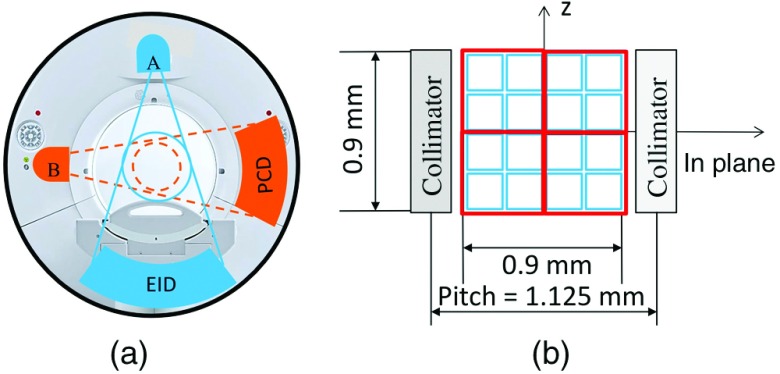
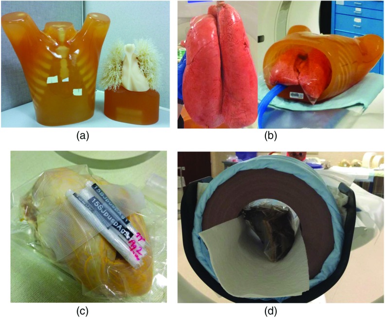
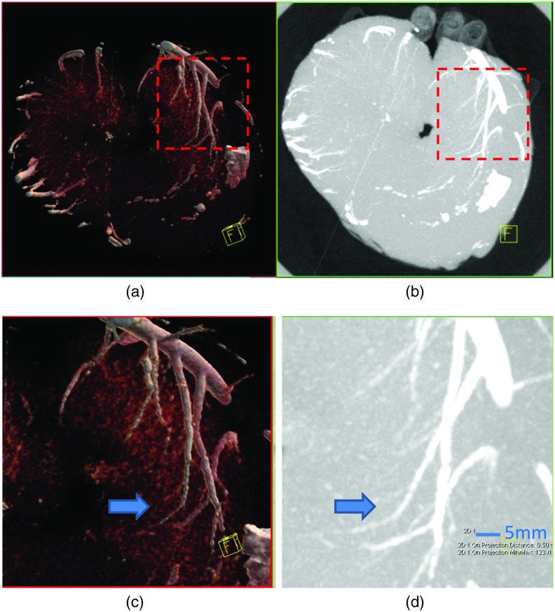
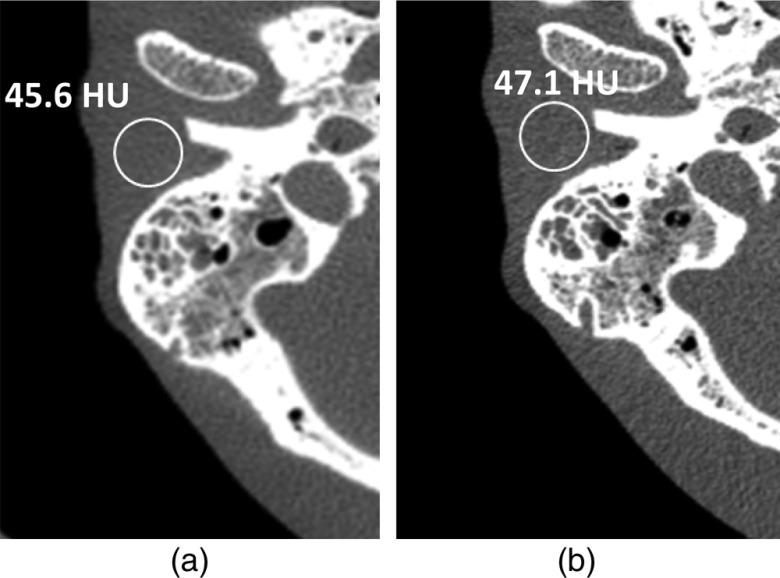
An ultrahigh-resolution (UHR) data collection mode was enabled on a whole-body, research photon counting detector (PCD) computed tomography system. In this mode, 64 rows of [Formula: see text] detector pixels were used, which corresponded to a pixel size of [Formula: see text] at the isocenter. Spatial resolution and image noise were quantitatively assessed for the UHR PCD scan mode, as well as for a commercially available UHR scan mode that uses an energy-integrating detector (EID) and a set of comb filters to decrease the effective detector size. Images of an anthropomorphic lung phantom, cadaveric swine lung, swine heart specimen, and cadaveric human temporal bone were qualitatively assessed. Nearly equivalent spatial resolution was demonstrated by the modulation transfer function measurements: 15.3 and [Formula: see text] spatial frequencies were achieved at 10% and 2% modulation, respectively, for the PCD system and 14.2 and [Formula: see text] for the EID system. Noise was 29% lower in the PCD UHR images compared to the EID UHR images, representing a potential dose savings of 50% for equivalent image noise. PCD UHR images from the anthropomorphic phantom and cadaveric specimens showed clear delineation of small structures.

Keywords: computed tomography; high-resolution mode; photon counting detector; spatial resolution.


Figures

Fig. 1
Fig. 1
(a) A research PCD-based CT system, built based on a second-generation dual-source CT system, consists of an EID and a PCD. (b) Detector configuration of the UHR mode of the PCD, showing both native pixels (blue) and UHR pixels (red).
Fig. 2
Fig. 2
A 50-μm diameter tungsten wire phantom (a) was placed in a 20 cm water tank (b) for MTF measurements.
Fig. 3
Fig. 3
Phantoms and specimens used in this study: (a) an anthropomorphic lung phantom with synthetic lung nodules; (b) a swine lung specimen inflated and placed in a hollow chest phantom; (c) a swine heart whose vessels were filled with lead-based Microfill®; and (d) a cadaveric temporal bone specimen placed in a 20 cm solid water phantom.
Fig. 4
Fig. 4
(a) MTF curves of the UHR mode demonstrate the same spatial resolution for head and body protocols, with a spatial resolution of 15.3 and 20.3  lp/cm at 10% and 2% modulation levels, respectively. (b) The MTF curve of the S80 kernel on the PCD system closely matched that of the modified S90 kernel on the EID system (14.2 and 18.6  lp/cm at 10% and 2% modulation levels, respectively).
Fig. 5
Fig. 5
(a, d) Axial and (b, c, e, f) volume rendered images of the anthropomorphic lung phantom scanned using the (a–c) UHR mode of the PCD subsystem and (d–f) UHR mode of the commercial EID system. The zoomed in ROIs (c, f) shows better delineation of submillimeter lung vessels using the PCD subsystem compared to the EID system.
Fig. 6
Fig. 6
(a) Axial and (b) MIP PCD UHR images of the right lung show clear boundaries of the low-contrast star-shaped lung nodule (arrow).
Fig. 7
Fig. 7
PCD UHR images of the swine lung at two different locations (a) and (b) show low attenuating parenchyma. Interlobular septa (arrow) and centrilobular ground-glass opacities (arrow head) surrounding small airways are clearly visible in the lung specimen image, especially within the dependent portions.
Fig. 8
Fig. 8
(a, c) Volume rendered and (b, d) MIP PCD UHR images of the heart specimen. Transmural coronary arteries (arrow), which are much smaller than 1 mm in diameter, are clearly visible in the images, especially with zoomed in ROIs (c, d).
Fig. 9
Fig. 9
Images of the temporal bone specimen scanned with (a) EID UHR and (b) PCD UHR modes. The malleus head and incus body are clearly visualized as an “ice cream cone” structure (arrow) on both images, but the PCD image demonstrates modest improvement in delineating the incudomallear joint (arrows). Improved sharpness and lower noise can be appreciated in the PCD image (arrow heads).
Fig. 10
Fig. 10
Images of the temporal bone specimen scanned with (a) EID UHR and (b) PCD UHR. Stapes superstructure was clearly delineated as a “wishbone” structure (arrow) on the PCD image, but was much less conspicuous on the EID image. Measurements of image noise were 59.6 HU for the EID image and 46.3 HU for the PCD image.
Fig. 11
Fig. 11
Images of the anthropomorphic phantom scanned on the EID (a) at full dose and PCD (b) at 50% dose. Measurements show image noise of 45.6 HU for the EID image at 0.6-mm slice thickness and 47.1 HU for the PCD image at 0.5-mm slice thickness.

References

    1. Hsieh J., Computed Tomography: Principles, Design, Artifacts, and Recent Advances, SPIE Press, Bellingham, Washington: (2003).
    1. Kalender W. A., Computed Tomography: Fundamentals, System Technology, Image Quality, Applications, 3rd ed., Publicis, Erlangen, Germany: (2011).
    1. McCollough C. H., et al. , “Spatial resolution improvement and dose reduction potential for inner ear CT imaging using a z-axis deconvolution technique,” Med. Phys. 40(6), 061904 (2013).10.1118/1.4802730 - DOI - PubMed
    1. Whiting B. R., et al. , “Use of computed tomography scans for cochlear implants,” J. Digital Imaging 21(3), 323–328 (2008).10.1007/s10278-007-9045-4 - DOI - PMC - PubMed
    1. Webb W. R., Muller N. L., Naidich D. P., High-Resolution CT of the Lung, 5th ed., Lippincott Williams & Wilkins, Philadelphia, Pennsylvania: (2014).