. 2016 Dec 31;22(1):62.
doi: 10.3390/molecules22010062.

A Novel Role of Silibinin as a Putative Epigenetic Modulator in Human Prostate Carcinoma

Affiliations

A Novel Role of Silibinin as a Putative Epigenetic Modulator in Human Prostate Carcinoma

Ioannis Anestopoulos et al. Molecules. .

Abstract

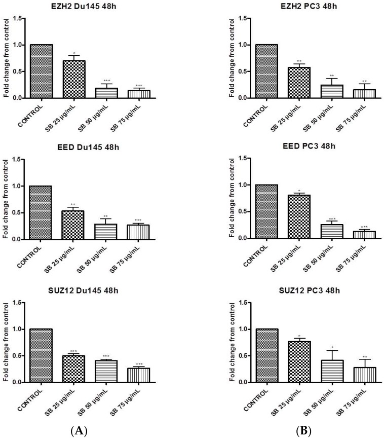
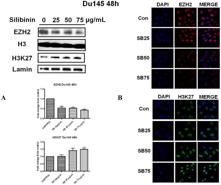
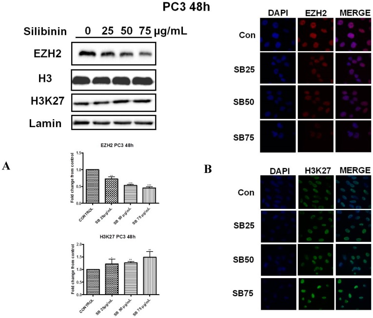
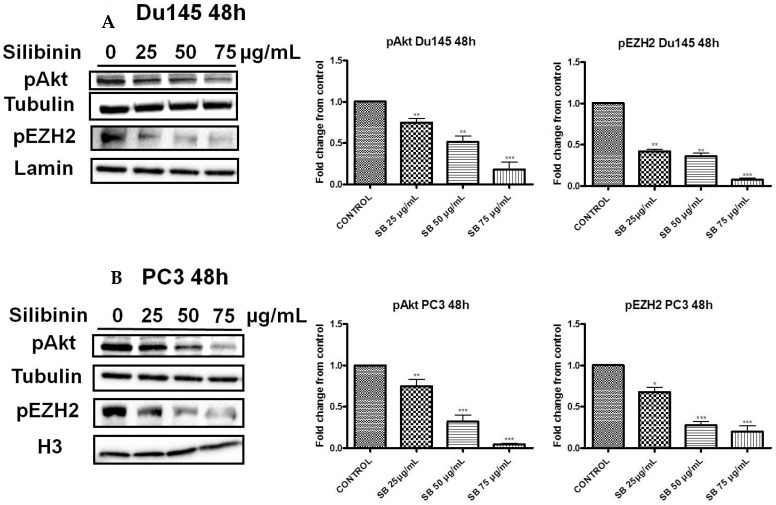
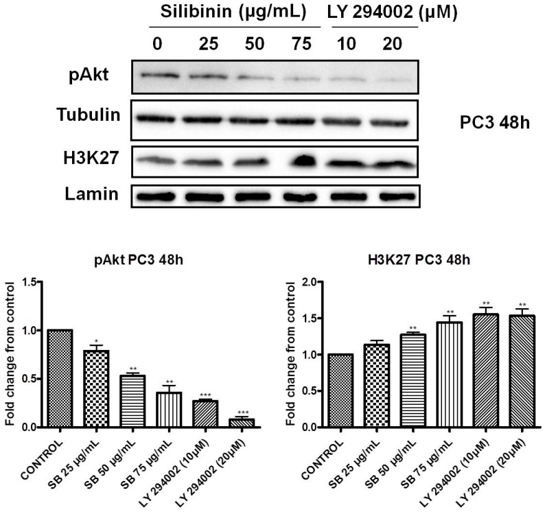
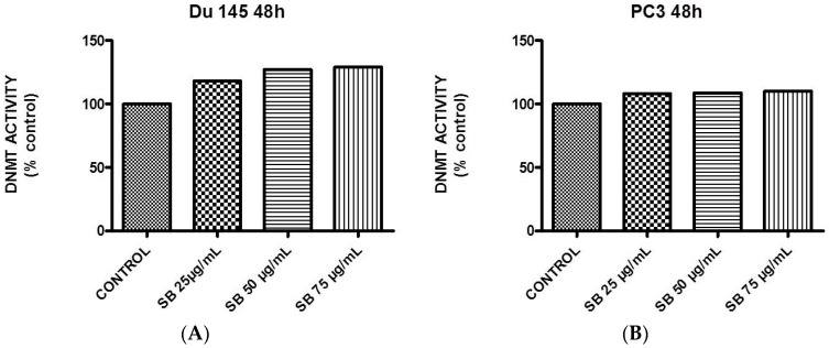
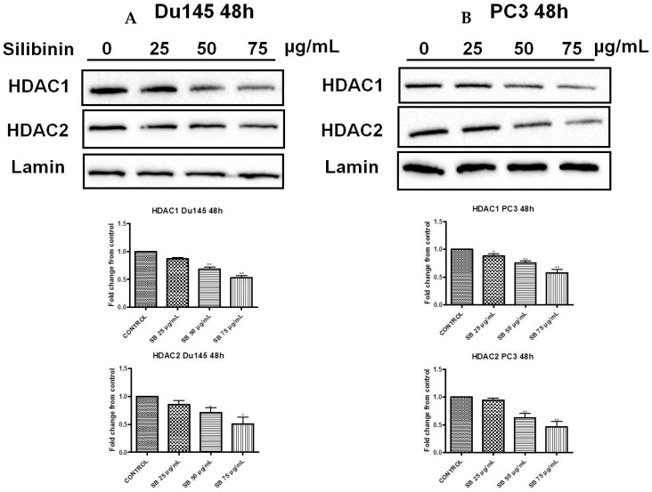
Silibinin, extracted from milk thistle (Silybum marianum L.), has exhibited considerable preclinical activity against prostate carcinoma. Its antitumor and chemopreventive activities have been associated with diverse effects on cell cycle, apoptosis, and receptor-dependent mitogenic signaling pathways. Here we hypothesized that silibinin's pleiotropic effects may reflect its interference with epigenetic mechanisms in human prostate cancer cells. More specifically, we have demonstrated that silibinin reduces gene expression levels of the Polycomb Repressive Complex 2 (PRC2) members Enhancer of Zeste Homolog 2 (EZH2), Suppressor of Zeste Homolog 12 (SUZ12), and Embryonic Ectoderm Development (EED) in DU145 and PC3 human prostate cancer cells, as evidenced by Real Time Polymerase Chain Reaction (RT-PCR). Furthermore immunoblot and immunofluorescence analysis revealed that silibinin-mediated reduction of EZH2 levels was accompanied by an increase in trimethylation of histone H3 on lysine (Κ)-27 residue (H3K27me3) levels and that such response was, in part, dependent on decreased expression levels of phosphorylated Akt (ser473) (pAkt) and phosphorylated EZH2 (ser21) (pEZH2). Additionally silibinin exerted other epigenetic effects involving an increase in total DNA methyltransferase (DNMT) activity while it decreased histone deacetylases 1-2 (HDACs1-2) expression levels. We conclude that silibinin induces epigenetic alterations in human prostate cancer cells, suggesting that subsequent disruptions of central processes in chromatin conformation may account for some of its diverse anticancer effects.

Keywords: DNMT; EZH2; H3K27me3; HDAC; PRC2; epigenetics; histone methylation; prostate cancer; silibinin.


Conflict of interest statement

The authors declare no conflict of interest.

Figures

Figure 1
Figure 1
Silibinin suppresses the expression of Polycomb Repressive Complex 2 (PRC2) complex members. DU145 (A) and PC3 (B) cells were incubated for 48h with different concentrations of silibinin (25–75 μg/mL). After treatments, cells were harvested for Real Time Polymerase Chain Reaction (RT-PCR) analysis of Enhancer of Zeste Homolog 2 (EZH2), Suppressor of Zeste Homolog 12 (SUZ12) and Embryonic Ectoderm Development (EED) genes. The expression levels of target genes were normalized to β-actin (internal control). The results are presented as mean ± SD of three independent experiments. * p < 0.05; ** p < 0.01; *** p < 0.001.
Figure 2
Figure 2
Silibinin reduces EZH2 expression while it increases trimethylation of Lys27 on H3 (H3K27me3). (A) DU145 cells were treated with different concentrations of silibinin (25–75 μg/mL) for 48 h. Nuclear extracts were analyzed for EZH2 and H3K27me3 by western blotting. Representative blots are shown. Equal protein loading was verified by stripping and re-probing the same membranes with histone H3 and lamin-β (2A upper panel). Quantification of EZH2 and H3K27me3 bands was performed by scanning densitometry (2A lower panel). The results are presented as the mean ± SD of three or more independent experiments. ** p < 0.01; *** p < 0.001; (B) DU145 cells were grown on coverslips in 6-well chamber plates for 24 h prior to treatments with various concentrations of silibinin (25–75 μg/mL) for 48 h. Cells were fixed and processed for staining with anti-EZH2 (2B upper panel) and anti-H3K27me3 (2B lower panel) antibodies, as described in ‘Materials and Methods’. Specific antibody staining was detected by fluorescein-conjugated secondary antibody and counterstained for DNA with 4′,6-diamidino-2-phenylindole (DAPI). The blue fluorescent staining column is for DAPI, the second red/green fluorescent column is for EZH2/H3K27me3 respectively, and the third column is for both images merged.
Figure 3
Figure 3
Silibinin reduces EZH2 expression while it increases trimethylation of Lys27 on H3 (H3K27me3). (A) PC3 cells were treated with different concentrations of silibinin (25–75 μg/mL) for 48 h. Nuclear extracts were analyzed for EZH2 and H3K27me3 by western blotting. Representative blots are shown. Equal protein loading was verified by stripping and re-probing the same membranes with histone H3 and lamin-β (3A upper panel). Quantification of EZH2 and H3K27me3 bands was performed by scanning densitometry (3A lower panel). The results are presented as the mean ± SD of three or more independent experiments. * p < 0.05; ** p < 0.01; *** p <0.001; (B) PC3 cells were grown on coverslips in 6-well chamber plates for 24h prior to treatments with different concentration of silibinin (25–75 μg/mL) for 48 h. Cells were fixed and processed for staining with anti-EZH2 (3B upper panel) and anti-H3K27me3 (3B lower panel) antibodies, as described in ‘Materials and Methods’. Specific antibody staining was detected by fluorescein-conjugated secondary antibody and counterstained for DNA with DAPI. The blue fluorescent staining column is for DAPI, the second red/green fluorescent column is for EZH2/H3K27me3, and the third column is for both images merged.
Figure 4
Figure 4
Silibinin decreases phosphorylation of pAkt (ser473) with a concomitant suppression of pEZH2 (ser21) levels. DU145 (A) and PC3 (B) cells were treated with different concentrations of silibinin (25–75 μg/mL) for 48 h. Cytosolic and nuclear extracts were analyzed for pAkt (ser473) and pEZH2 (ser21) by western blotting, respectively. Representative blots are shown. Equal protein loading was verified by stripping and re-probing the same membranes with tubulin and histone H3/lamin-β (left panel). Quantification of pAkt and pEZH2 bands was performed by scanning densitometry (right panel). The results are presented as the mean ± SD of three or more independent experiments. * p < 0.05; ** p < 0.01; *** p < 0.001.
Figure 5
Figure 5
Increased trimethylation of lysine 27 on histone H3 by silibinin is associated with decreased pAkt levels. PC3 cells were treated with different concentrations of silibinin (25–75 μg/mL) and PI3K inhibitor LY294002 (10–20 μΜ) for 48 h. Cytosolic and nuclear extracts were analyzed for pAkt (ser473) and H3K27me3 by western blotting, respectively. Representative blots are shown. Equal protein loading was verified by stripping and re-probing the same membranes with tubulin and lamin-β (upper panel). Quantification of pAkt (ser473) and H3K27me3 bands was performed by scanning densitometry (lower panel). The results are presented as the mean ± SD of three or more independent experiments. * p < 0.05, ** p < 0.01, *** p < 0.001.
Figure 6
Figure 6
Silibinin induces a concentration-dependent increase in DNA methyltransferase activity. Logarithmically-growing DU145 (A) and PC3 (B) cells were treated with 25–75 μg/mL silibinin for 48 h and processed for quantification of global DNA methyltransferase activity.
Figure 7
Figure 7
Silibinin induces a concentration-dependent decrease of HDAC1-2 expression levels. DU145 (A) and PC3 (B) cells were treated with different concentrations of silibinin (25–75 μg/mL) for 48 h. Nuclear extracts were analyzed for HDAC1 and HDAC2 by western blotting. Representative blots are shown. Equal protein loading was verified by stripping and re-probing the same membranes with lamin-β (upper panel). Quantification of HDACs1-2 bands was performed by scanning densitometry (lower panel). The results are presented as the mean ± SD of three or more independent experiments. * p < 0.05; ** p < 0.01.

References

    1. Ferlay J., Shin H.R., Bray F., Forman D., Mathers C., Parkin D.M. Estimates of worldwide burden of cancer in 2008: GLOBOCAN 2008. Int. J. Cancer. 2010;127:2893–2917. doi: 10.1002/ijc.25516. - DOI - PubMed
    1. Jemal A., Bray F., Center M.M., Ferlay J., Ward E., Forman D. Global cancer statistics. CA Cancer J. Clin. 2011;61:69–90. doi: 10.3322/caac.20107. - DOI - PubMed
    1. Chiam K., Ricciardelli C., Bianco-Miotto T. Epigenetic biomarkers in prostate cancer: Current and future uses. Cancer Lett. 2014;342:248–256. doi: 10.1016/j.canlet.2012.02.011. - DOI - PubMed
    1. Chin S.P., Dickinson J.L., Holloway A.F. Epigenetic regulation of prostate cancer. Clin. Epigenet. 2011;2:151–169. doi: 10.1007/s13148-011-0041-7. - DOI - PMC - PubMed
    1. Kallifatidis G., Hoy J.J., Lokeshwar B.L. Bioactive natural products for chemoprevention and treatment of castration-resistant prostate cancer. Semin. Cancer Biol. 2016;40–41:160–169. doi: 10.1016/j.semcancer.2016.06.003. - DOI - PMC - PubMed

MeSH terms

LinkOut - more resources