

Targeting antioxidant enzyme expression as a therapeutic strategy for ischemic stroke
- PMID: 28043837
- PMCID: PMC5461189
- DOI: 10.1016/j.neuint.2016.12.007
Targeting antioxidant enzyme expression as a therapeutic strategy for ischemic stroke
Abstract
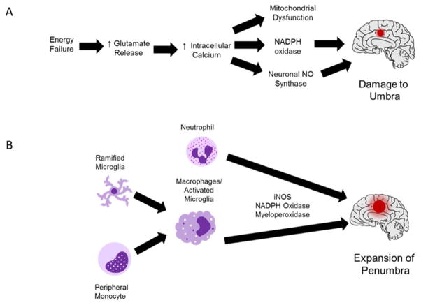
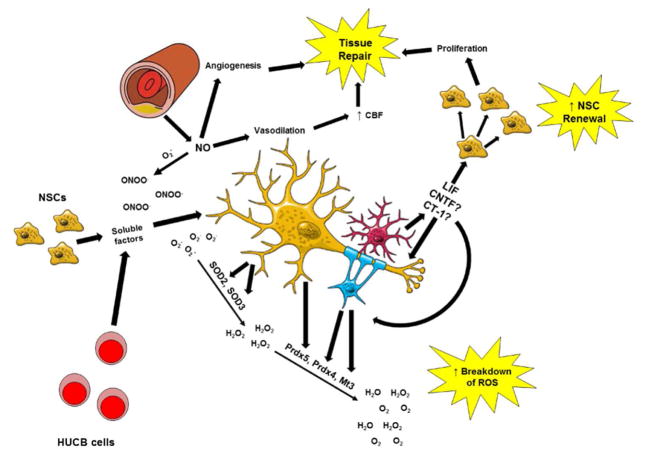
During ischemic stroke, neurons and glia are subjected to damage during the acute and neuroinflammatory phases of injury. Production of reactive oxygen species (ROS) from calcium dysregulation in neural cells and the invasion of activated immune cells are responsible for stroke-induced neurodegeneration. Scientists have failed thus far to identify antioxidant-based drugs that can enhance neural cell survival and improve recovery after stroke. However, several groups have demonstrated success in protecting against stroke by increasing expression of antioxidant enzymes in neural cells. These enzymes, which include but are not limited to enzymes in the glutathione peroxidase, catalase, and superoxide dismutase families, degrade ROS that otherwise damage cellular components such as DNA, proteins, and lipids. Several groups have identified cellular therapies including neural stem cells and human umbilical cord blood cells, which exert neuroprotective and oligoprotective effects through the release of pro-survival factors that activate PI3K/Akt signaling to upregulation of antioxidant enzymes. Other studies demonstrate that treatment with soluble factors released by these cells yield similar changes in enzyme expression after stroke. Treatment with the cytokine leukemia inhibitory factor increases the expression of peroxiredoxin IV and metallothionein III in glia and boosts expression of superoxide dismutase 3 in neurons. Through cell-specific upregulation of these enzymes, LIF and other Akt-inducing factors have the potential to protect multiple cell types against damage from ROS during the early and late phases of ischemic damage.
Keywords: Antioxidant enzymes; Ischemic stroke; Leukemia inhibitory factor; Neuroprotection; Oxidative stress.
Copyright © 2016 Elsevier Ltd. All rights reserved.
Conflict of interest statement
Figures
References
-
- Aksenov MY, Tucker HM, Nair P, Aksenova MV, Butterfield DA, Estus S, Markesbery WR. The expression of key oxidative stress-handling genes in different brain regions in Alzheimer’s disease. J Mol Neurosci. 1998;11:151–164. - PubMed
-
- Alonzi T, Middleton G, Wyatt S, Buchman V, Betz AKU, Muller W, Musiani P, Poli V, Davies AM. Role of STAT3 and PI 3-Kinase/Akt in Mediating the Survival Actions of Cytokines on Sensory Neurons. Molecular and Cellular Neuroscience. 2001;18:270–282. - PubMed
-
- Antonyuk SV, Strange RW, Marklund SL, Hasnain SS. The structure of human extracellular copper–zinc superoxide dismutase at 1.7 Å resolution: insights into heparin and collagen binding. J Mol Bio. 2009;388:310–326. - PubMed
-
- Arce V, Garces A, de Bovis B, Filippi P, Henderson C, Pettmann B, deLapeyrière O. Cardiotrophin-1 requires LIFRβ to promote survival of mouse motoneurons purified by a novel technique. J Neurosci Res. 1999;55:119–126. - PubMed
-
- Arthan D, Hong SK, Park JI. Leukemia inhibitory factor can mediate Ras/Raf/MEK/ERK-induced growth inhibitory signaling in medullary thyroid cancer cells. Cancer Letters. 2010;297:31–41. - PubMed
Publication types
MeSH terms
Substances
Grants and funding
LinkOut - more resources
Full Text Sources
Other Literature Sources
Medical
Research Materials
Miscellaneous
