. 2017 Jul;140(1):109-120.
doi: 10.1016/j.jaci.2016.08.056. Epub 2016 Dec 31.

IL-1 and IL-36 are dominant cytokines in generalized pustular psoriasis

Affiliations

IL-1 and IL-36 are dominant cytokines in generalized pustular psoriasis

Andrew Johnston et al. J Allergy Clin Immunol. 2017 Jul.

Abstract

Background: Generalized pustular psoriasis (GPP) is a rare, debilitating, and often life-threatening inflammatory disease characterized by episodic infiltration of neutrophils into the skin, pustule development, and systemic inflammation, which can manifest in the presence or absence of chronic plaque psoriasis (PV). Current treatments are unsatisfactory and warrant a better understanding of GPP pathogenesis.

Objective: We sought to understand better the disease mechanism of GPP to allow improved targeted therapies.

Methods: We performed a gene expression study on formalin-fixed paraffin-embedded GPP (n = 28) and PV (n = 12) lesional biopsies and healthy control (n = 20) skin. Differential gene expression was analyzed using gene ontology and enrichment analysis. Gene expression was validated with quantitative RT-PCR and immunohistochemistry, and a potential disease mechanism was investigated using primary human cell culture.

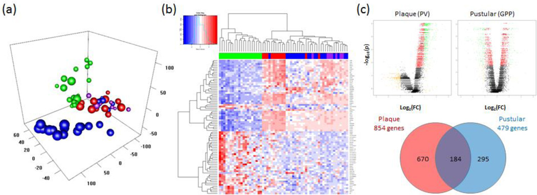
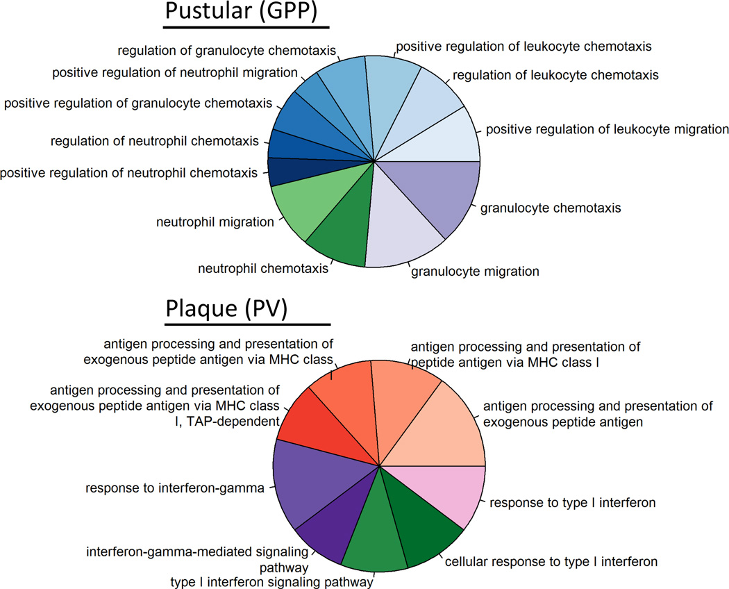
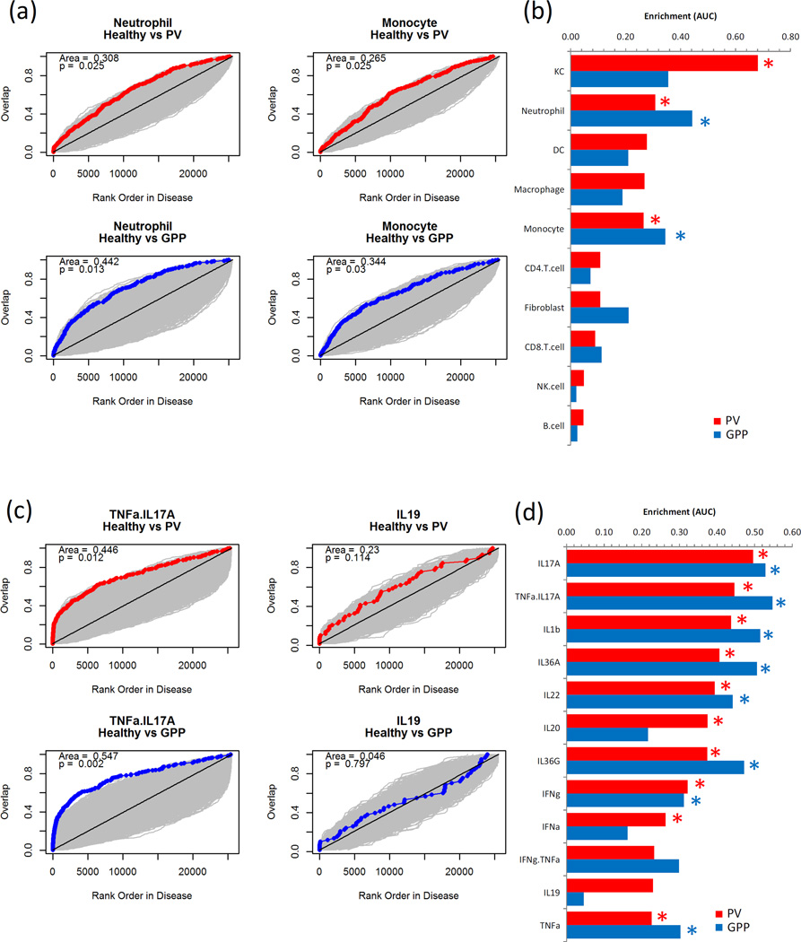
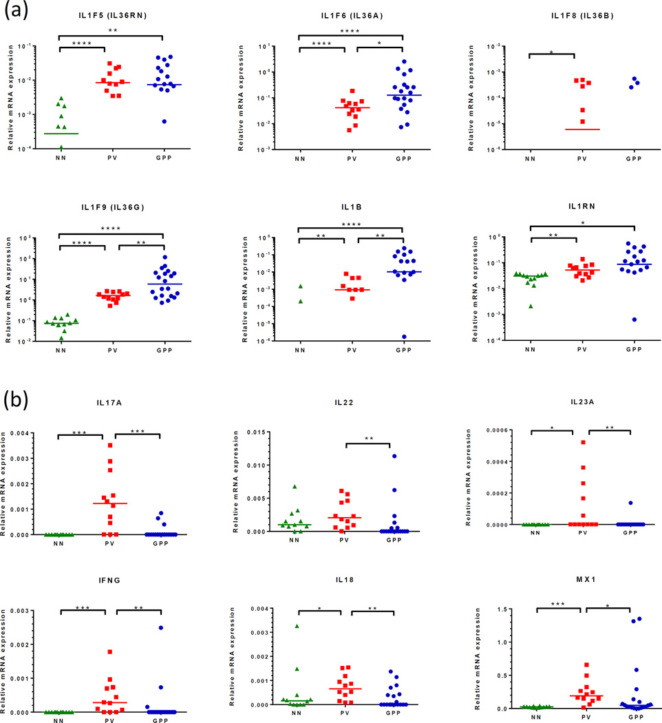
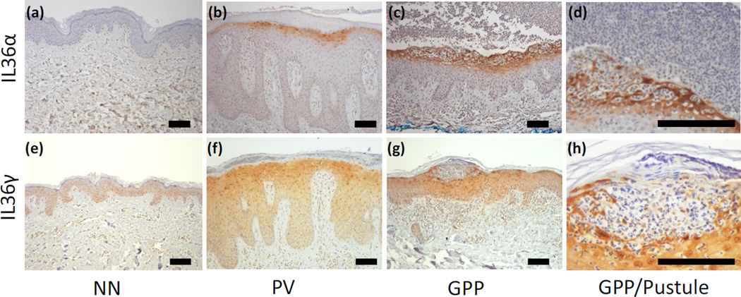
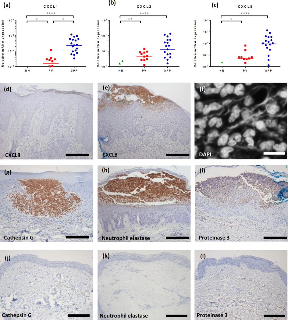
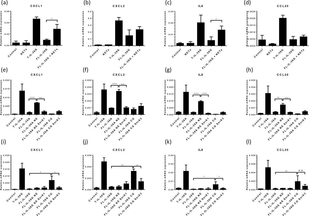
Results: Compared with healthy skin, GPP lesions yielded 479 and PV 854 differentially expressed genes, respectively, with 184 upregulated in both diseases. We detected significant contributions of IL-17A, TNF, IL-1, IL-36, and interferons in both diseases; although GPP lesions furnished higher IL-1 and IL-36 and lower IL-17A and IFN-γ mRNA expression than PV lesions did. We detected prominent IL-36 expression by keratinocytes proximal to neutrophilic pustules, and we show that both neutrophils and neutrophil proteases activate IL-36. Suggesting another mechanism regulating IL-36 activity, the protease inhibitors serpin A1 and A3, which inhibit elastase and cathepsin G, respectively, were upregulated in both diseases and inhibited activation of IL-36.

Conclusions: Our data indicate sustained activation of IL-1 and IL-36 in GPP, inducing neutrophil chemokine expression, infiltration, and pustule formation, suggesting that the IL-1/IL-36 inflammatory axis is a potent driver of disease pathology in GPP.

Keywords: Generalized pustular psoriasis; inflammation; interleukin; psoriasis.


Figures

Figure 1
Figure 1. The transcriptome of generalized pustular psoriasis shares features of plaque psoriasis but is skewed towards innate immune inflammation
Formalin-fixed paraffin-embedded biopsies of chronic plaque psoriasis (PV, n=12), generalized pustular psoriasis (GPP, n=28) and healthy control skin (NN, n=20) were processed for RNA extraction and analyzed using Affymetrix ST 2.1 arrays. Principal component analysis of the GPP, PV and NN samples showed segregation of the GPP and PV samples from the healthy samples, with some overlap between the plaque and pustular samples. NN, PV, PV+GPP and GPP-only phenotypes are indicated by green, red, purple and blue spheres respectively (a). A heat map generated from unsupervised clustering of differentially expressed transcripts between GPP, PV and NN skin shows heterogeneity of healthy and disease samples. For clarity, the 79 transcripts with 0.25>FC>4.0, and FDR p<0.05 were selected. Blue indicates low expression levels whereas red indicates high expression levels. The green, red, purple and blue bars above the heat map indicate NN, PV, PV+GPP and GPP-only skin samples respectively (b). Volcano plots of gene expression showing roughly equal numbers of up- and down-regulated genes in each disease although many of the down-regulated transcripts in PV did not reach statistical significance. Yellow indicates transcripts with 0.5>FC>2, red denotes FDR p<0.05, and green labels transcripts satisfying both criteria (upper panels, c). Despite overlap of differentially expressed genes between PV and GPP skin lesions, each disease had a set of uniquely-expressed transcripts (lower panel, c).
Figure 2
Figure 2. The transcriptome of pustular psoriasis lesions are enriched for innate immune genes
Gene ontology analysis of DEGs in GPP and PV using the GO_ImmuneSystemProcess category. GO analysis highlighted an over-representation of innate immune genes in GPP (A) contrasting with more prominent expression of genes involved in acquired immunity in PV (B). Categories shown are those reaching statistical significance using a two-sided hypergeometric test. Details of individual GO terms are listed in Supplemental Table 2.
Figure 3
Figure 3. Neutrophil and monocyte transcripts show greater enrichment in pustular psoriasis (GPP) lesions than plaque psoriasis (PV), whereas robust cytokine gene induction is evident in both diseases
Microarray datasets for KC, CD4+ T-cells, CD8+ T-cells, monocytes, neutrophils and dendritic cells (DC) were compiled and used to generate signature gene lists (13). DEGs in PV vs NN and GPP vs NN comparisons were ordered by fold-change and the rank position each of the cell signature genes noted and cumulative overlap between the sets plotted (red/blue line), showing a greater up-regulation of neutrophil-specific genes in GPP than PV lesions. Grey areas indicate space generated by 1000 random permutations of NN, PV and GPP datasets from which statistical significance is derived (a). Data from each of the cell signature comparisons is summarized, red bars PV, blue bars GPP, statistical significance indicated by *p<0.05 permutation test (b). Likewise gene signatures for cytokine activity on KC were generated and used to discern cytokine activity within the PV and GPP samples, showing prominent and significant presence of genes induced by TNF-α+IL17A in PV and GPP lesions, contrasted by the lack of enrichment in genes induced by IL-19 (c and d).
Figure 4
Figure 4. GPP lesions have a heightened IL-1/IL-36 cytokine axis but less pronounced Th1/Th17 gene expression than PV
Quantitative RT-PCR revealed more abundant expression of IL36A, IL36G and IL1B transcripts in GPP compared with PV biopsies, this difference was not seen with respect to the receptor antagonists IL1RN and IL36RN (a). Suggesting a departure from typical Th1/Th17 pathophysiology, IL23A, IL17A, IFNG, CXCL9, CXCL10 and MX1 transcript expression was found to be significantly lower in GPP compared to PV lesions (b). Statistical significance indicated *p<0.05, **p<0.01, ***p<0.001, ****p<0.0001. Kruskal-Wallace test with Dunn’s multiple comparison test.
Figure 5
Figure 5. Immunohistochemical detection of IL-36α and IL-36γ in skin confirms elevated expression of IL-36 in GPP and localizes IL-36 expression to keratinocytes surrounding the neutrophilic pustule
IL-36α was not expressed by healthy skin (a), found in the cytoplasm of the uppermost layers of viable KC in PV lesions (b) and expressed at a higher intensity by a wider band of KC in GPP lesions (c). IL-36α expression was strongest in the LC proximal to neutrophilic pustules, but not the neutrophils themselves (d). Faint expression of IL-36γ could be detected in the cytoplasm, but was most apparent in the nuclei of healthy skin KC (e). IL-36γ was intensified in PV lesions (f) yet this was more intense in GPP lesions (g), most pronounced in the KC adjacent to pustules (h). Representative images from 6 GPP, PV and NN donors used (Supplemental Figure 1). DAB with hematoxylin counterstain. Scale bar 100µm.
Figure 6
Figure 6. The neutrophil chemokines CXCL1, CXCL2 and CXCL8 are more strongly expressed in GPP than PV lesions
Using qRT-PCR we detected significantly higher expression of CXCL1, CXCL2, and IL8 transcripts in GPP (n=20) compared with plaque psoriasis (n=12) or healthy control skin (NN, n=12) (a–c). CXCL8 immunoreactivity was barely evident in PV (d) but very strong in and around neutrophilic pustules in GPP biopsies (e). NET formation was prominent in GPP lesions as visualized with DAPI (f) as was the expression of the neutrophil proteases cathepsin G, elastase, and proteinase 3 in GPP (g, h, i) but not healthy control skin (j, k, l). Representative images of at least 6 GPP, PV, NN donors used (Supplemental Figure 1). Scale bar, d- e, and g-l: 200µm, f: 10µm. Statistical significance indicated *p<0.05, **p<0.01, ***p<0.001, ****p<0.0001. Kruskal-Wallace test with Dunn’s multiple comparison test.
Figure 7
Figure 7. Neutrophil extracellular traps (NETs) and neutrophil proteases activate IL-36
Exposure to NETs for 1 hour (a–d) significantly increased the ability of full-length (FL)-IL-36γ to induce CXCL1 and IL8 expression by KC more than 3-fold compared with untreated FL-IL-36γ (a and c), suggesting that enzyme activity within the NETs caused FL-IL-36γ activation. Truncated (T)-IL-36 as positive control for IL-36 activity. Purified neutrophil elastase (NE, e–h) and cathepsin G (CG, i–l) activated FL-IL-36α and FL-IL-36γ respectively which was inhibited by the addition of specific inhibitors of elastase (serpin A1) or cathepsin G (serpin A3). Statistical significance determined with Student’s t-test and indicated *p<0.05, **p<0.01, ***p<0.001, ****p<0.0001, n.s. non-significant.

References

    1. Gudjonsson JE, Elder JT. Chapter 18. Psoriasis. In: Goldsmith LA, Katz SI, Gilchrest BA, Paller AS, Leffell DJKW, editors. Fitzpatrick’s dermatology in general medicine. 1. 8th. New York: McGraw-Hill Medical; 2012.
    1. Hussain S, Berki DM, Choon SE, Burden AD, Allen MH, Arostegui JI, et al. IL36RN mutations define a severe autoinflammatory phenotype of generalized pustular psoriasis. J Allergy Clin Immunol. 2015;135(4):1067–1070. e9. - PubMed
    1. Onoufriadis A, Simpson MA, Pink AE, Di Meglio P, Smith CH, Pullabhatla V, et al. Mutations in IL36RN/IL1F5 are associated with the severe episodic inflammatory skin disease known as generalized pustular psoriasis. Am J Hum Genet. 2011;89(3):432–437. - PMC - PubMed
    1. Setta-Kaffetzi N, Navarini AA, Patel VM, Pullabhatla V, Pink AE, Choon SE, et al. Rare pathogenic variants in IL36RN underlie a spectrum of psoriasis-associated pustular phenotypes. J Invest Dermatol. 2013;133(5):1366–1369. - PubMed
    1. Sugiura K, Takemoto A, Yamaguchi M, Takahashi H, Shoda Y, Mitsuma T, et al. The majority of generalized pustular psoriasis without psoriasis vulgaris is caused by deficiency of interleukin-36 receptor antagonist. J Invest Dermatol. 2013;133(11):2514–2521. - PubMed