. 2017 Jan:57-58:244-257.
doi: 10.1016/j.matbio.2016.12.003. Epub 2016 Dec 30.

Integrin alpha6 maintains the structural integrity of the kidney collecting system

Affiliations

Integrin alpha6 maintains the structural integrity of the kidney collecting system

Olga M Viquez et al. Matrix Biol. 2017 Jan.

Abstract

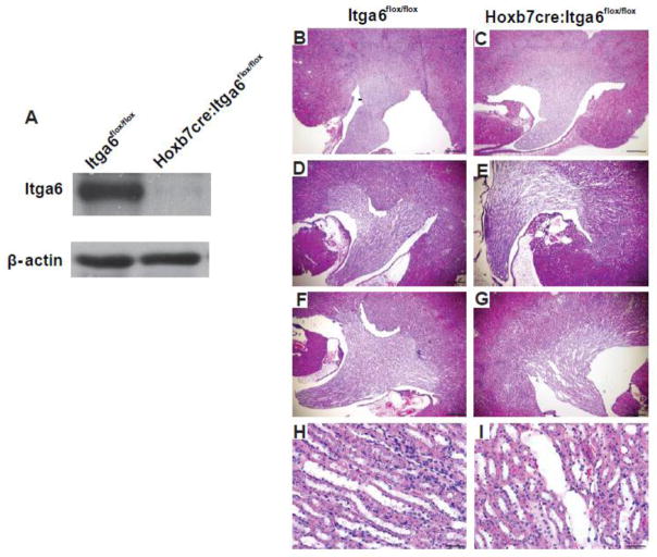
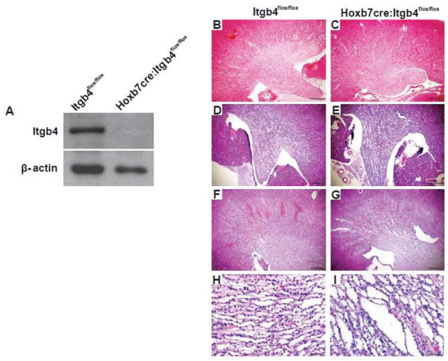
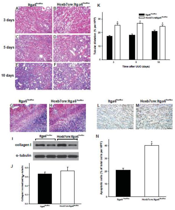
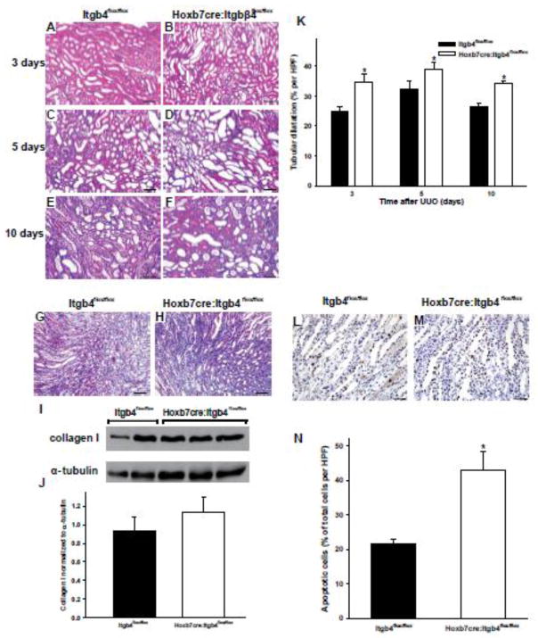
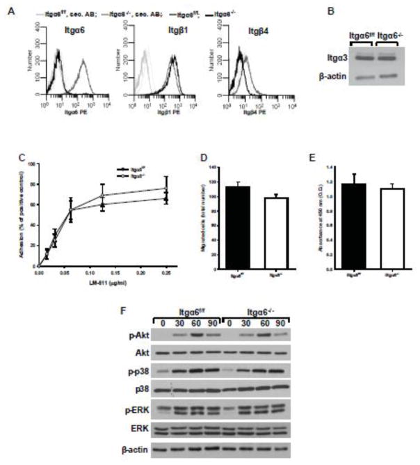
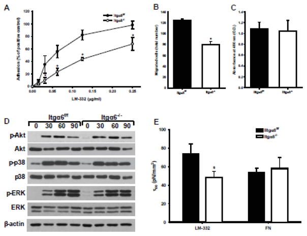
Laminins are a major constituent of the basement membranes of the kidney collecting system. Integrins, transmembrane receptors formed by non-covalently bound α and β subunits, serve as laminin receptors, but their role in development and homeostasis of the kidney collecting system is poorly defined. Integrin α3β1, one of the major laminin receptors, plays a minor role in kidney collecting system development, while the role of α6 containing integrins (α6β1 and α6β4), the other major laminin receptors, is unknown. Patients with mutations in α6 containing integrins not only develop epidermolysis bullosa, but also have abnormalities in the kidney collecting system. In this study, we show that selectively deleting the α6 or β4 integrin subunits at the initiation of ureteric bud development in mice does not affect morphogenesis. However, the collecting system becomes dilated and dysmorphic as the mice age. The collecting system in both null genotypes was also highly susceptible to unilateral ureteric obstruction injury with evidence of excessive tubule dilatation and epithelial cell apoptosis. Mechanistically, integrin α6-null collecting duct cells are unable to withstand high mechanical force when adhered to laminin. Thus, we conclude that α6 integrins are important for maintaining the integrity of the kidney collecting system by enhancing tight adhesion of the epithelial cells to the basement membrane. These data give a mechanistic explanation for the association between kidney collecting system abnormalities in patients and epidermolysis bullosa.


Conflict of interest statement

The authors declare no competing or financial interests.

Figures

Figure 1
Figure 1. The integrin α6 subunit is not required for normal UB morphogenesis but is necessary to maintain normal kidney collecting duct morphology
(A) Expression of integrin α6 in papillary lysates from a 3-day old Itga6flox/flox and Hoxb7cre:Itga6flox/flox mice was analyzed by Western blotting analysis. α6 is indicated by the arrowhead. (B–I) Morphology of H&E stained kidney sections from Itga6flox/flox and Hoxb7cre:Itga6flox/flox mice at 2 months (magnification 40×, scale bar =500um) (B, C), 6 months (magnification 40×, scale bar =500um), (D, E) and 10 months of age (F, G) (magnification 40×, scale bar =500um) and (H, I) (magnification 200×, scale bar =100um).
Figure 2
Figure 2. The integrin β4 subunit is not required for normal UB morphogenesis but is necessary to maintain normal kidney collecting duct morphology
A) Expression of integrin β4 in papillary lysates from a 3-day old Itgb4flox/flox and Hoxb7cre:Itgb4flox/flox mice analyzed by Western blotting analysis. (B–I) Morphology of H&E stained kidney sections from Itgb4flox/flox and Hoxb7cre:Itgb4flox/flox mice at 2 months (magnification 40×, scale bar =500um) (B, C), 6 months (magnification 40×, scale bar =500um), (D, E) and 12 months of age (F, G) (magnification 40×, scale bar =500um) and (H, I) (magnification 200×, scale bar =100um).
Figure 3
Figure 3. Hoxb7cre:Itga6flox/flox mice have increased injury after UUO
(A–F) Morphology of H&E stained kidney sections from Itga6flox/flox (A, C, E) and Hoxb7cre:Itga6flox/flox (B, D, F) 2 month old mice subjected to UUO for 3 (A, B), 5 (C, D) or 10 (E, F) days (magnification 200×, scale bar =100um). (G, H) Fibrillar collagen was detected 7 days after UUO by Trichrome staining. (I, J) Levels of collagen I in kidney lysates of Itga6flox/flox and Hoxb7cre:Itga6flox/flox mice at 7 days after UUO was assessed by Western analysis and quantified by densitometry, normalized to α-tubulin and reported as mean measurements ±SEM. (K) Quantitative analysis of tubular dilatation in renal sections of Itga6flox/flox (black bars) and Hoxb7cre:Itga6flox/flox (white bars) mice subjected to UUO for 3, 5 or 10 days. Values are means with SEM from 6 mice; *p<0.01between Itga6flox/flox and Hoxb7cre:Itga6flox/flox mice. (L, M) Kidney papilla sections from UUO-treated Itga6flox/flox and Hoxb7cre:Itga6flox/flox mice for 2 days were stained for TUNEL (apoptosis) (scale bar= 100um). (N) Quantitative analysis of apoptosis in renal papilla sections of UUO-treated Itga6flox/flox and Hoxb7cre:Itga6flox/flox mice. Apoptosis was quantified and expressed as percent of apoptotic cells per microscopic field (9 fields of 9 kidneys from either genotype were analyzed. *p<0.01 between Itga6flox/flox and Hoxb7cre:Itga6flox/flox mice.
Figure 4
Figure 4. Hoxb7cre:Itgb4flox/flox mice have increased injury after UUO
(A–F) Morphology of H&E stained kidney sections from Itgb4flox/flox (A, C, E) and Hoxb7cre:Itgb4flox/flox (B, D, F) 2 month old mice subjected to UUO for 3 (A, B), 5 (C, D) or 10 (E, F) days (magnification 200×, scale bar =100um). (G, H) Fibrillar collagen was detected 7 days after UUO by Trichrome staining. (I, J) Levels of collagen I in kidney lysates of Itgb4flox/flox and Hoxb7cre:Itgb4flox/flox mice at 7 days after UUO was assessed by Western analysis and quantified by densitometry, normalized to α-tubulin and reported as mean measurements ±SEM. (K) Quantitative analysis of tubular dilatation in renal sections of Itgb4flox/flox (black bars) and Hoxb7: Itgβ4flox/flox (white bars) mice subjected to UUO for 3, 5 or 10 days. Values are means with SEM from 6 mice;a significant *p<0.01 between Itgb4flox/flox and Hoxb7cre:Itgb4flox/flox mice. (L, M) Kidney sections from UUO-treated Itgb4flox/flox and Hoxb7cre:Itgb4flox/flox mice for 2 days were stained for TUNEL (apoptosis)( scale bar =100um). (N) Quantitative analysis of apoptosis in renal sections of UUO-treated Itgb4flox/flox and Hoxb7cre:Itgb4flox/flox mice. Apoptosis was quantified and expressed as percent of apoptotic cells per microscopic field (9 fields of 9 kidneys from either genotype were analyzed *p<0.01 between Itgb4flox/flox and Hoxb7cre:Itgb4flox/flox mice.
Figure 5
Figure 5. Itgα6−/− CD cells adhere, migrate, proliferate and signal normally on LM-511
(A) Surface expression of integrin α6, β1 and β4 subunits were determined on Itgα6f/f and Itgα6−/− CD cells by flow cytometry using R-phycoerythrin (PE) conjugated secondary antibodies. (B) Lysates from Itgα6f/f and Itgα6−/− CD cells (20 μg total protein/lane) were immunoblotted for integrin α3 subunits or β-actin (loading control). Adhesion (C), migration (D) and proliferation (E) on LM-511 were evaluated as described in the Methods. For migration and proliferation, 1 μg/ml LM-511 was used. Shown are mean measurements ±SEM of 4–6 independent experiments. (F) Itgα6f/f and Itgα6−/− CD cells were plated in serum-free medium on LM-511 (1 μg/ml). Cells were lysed at 30, 60 and 90 min after plating and lysates (20 μg total protein/lane) were analyzed by Western blot for levels of phosphorylated and total Akt, p38, and ERK1/2 or β-actin (loading control).
Figure 6
Figure 6. Itgα6−/− CD cells show adhesion, migration and adhesion strength defects on LM-332
Adhesion (A), migration (B), proliferation (C), and replating assay (D) on LM-322 (1 μg/ml) were evaluated as described in the Methods and in Fig. 5. Shown are mean measurements ±SEM of 4–6 independent experiments; *p<0.01between Itgα6−/− and Itgα6f/f CD cells. (E) The shear stress for 50% detachment (τ50), which represents the mean cell adhesion strength, was determined for Itgα6−/− and Itgα6f/f CD cells on LM-332 and fibronectin (which is not a ligand of a6 containing integrin) coated cell culture plates. Shown are the averaged τ50 values of 3 independent experiments; *p<0.01between Itgα6−/− and Itgα6f/f CD cells.

References

    1. Mathew S, Chen X, Pozzi A, Zent R. Integrins in renal development. Pediatric nephrology. 2012;27(6):891–900. - PubMed
    1. Pozzi A, Zent R. Extracellular matrix receptors in branched organs. Curr Opin Cell Biol. 2011;23(5):547–53. - PMC - PubMed
    1. Pozzi A, Zent R. Integrins in kidney disease. Journal of the American Society of Nephrology : JASN. 2013;24(7):1034–9. - PMC - PubMed
    1. Pozzi A, Zent R. Integrins: sensors of extracellular matrix and modulators of cell function. Nephron Exp Nephrol. 2003;94(3):e77–84. - PubMed
    1. Miner JH, Yurchenco PD. Laminin functions in tissue morphogenesis. Annu Rev Cell Dev Biol. 2004;20:255–84. - PubMed

Publication types

MeSH terms

LinkOut - more resources