Review
. 2017 Feb;216(2):305-315.
doi: 10.1083/jcb.201610042. Epub 2017 Jan 2.

Mechanosensing by the nucleus: From pathways to scaling relationships

Affiliations
Review

Mechanosensing by the nucleus: From pathways to scaling relationships

Sangkyun Cho et al. J Cell Biol. 2017 Feb.

Abstract

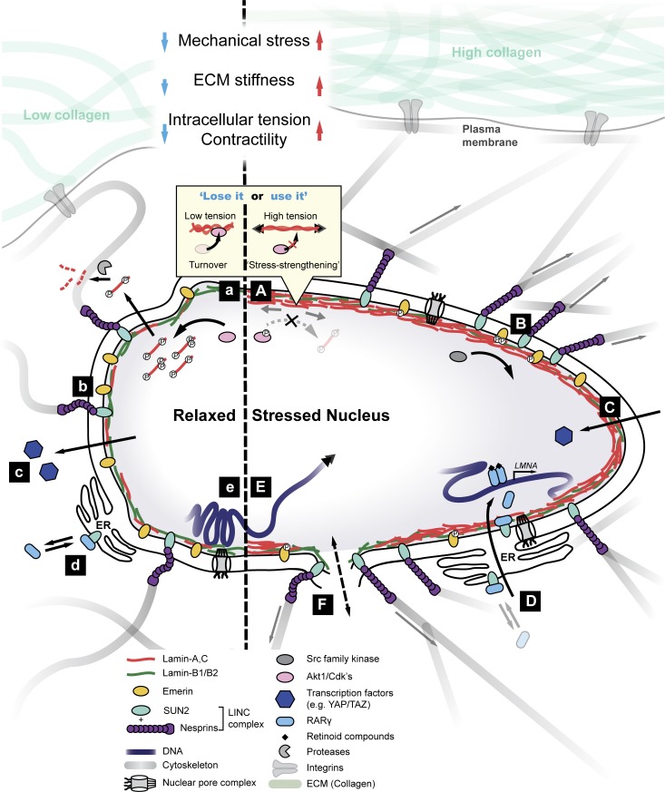
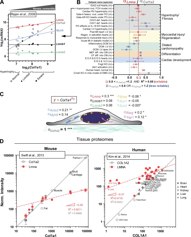
The nucleus is linked mechanically to the extracellular matrix via multiple polymers that transmit forces to the nuclear envelope and into the nuclear interior. Here, we review some of the emerging mechanisms of nuclear mechanosensing, which range from changes in protein conformation and transcription factor localization to chromosome reorganization and membrane dilation up to rupture. Nuclear mechanosensing encompasses biophysically complex pathways that often converge on the main structural proteins of the nucleus, the lamins. We also perform meta-analyses of public transcriptomics and proteomics data, which indicate that some of the mechanosensing pathways relaying signals from the collagen matrix to the nucleus apply to a broad range of species, tissues, and diseases.


Figures

Figure 1.
Figure 1.
Nucleus mechanosensation. Left and right sides indicate relaxed (soft) and mechanically stressed nuclei, respectively. (A and a) High nuclear tension can induce conformational changes in lamin coiled-coil dimers, which sterically inhibits access by kinases (Swift et al., 2013; Buxboim et al., 2014). In a relaxed nucleus, lamins are more phosphorylated and solubilized into the nucleoplasm (as during cell division). Phospho-solubilized lamins may ultimately become degraded (Bertacchini et al., 2013; Buxboim et al., 2014). Tension-inhibited turnover of lamins is similar to that of collagen I (Flynn et al., 2010) and is an example of structural proteins exhibiting stress-strengthening properties. (B and b) Pulling on nesprin-1 leads to phosphorylation of emerin by Src kinases (Guilluy et al., 2014) and results in stress stiffening of the nucleus. Emerin phosphorylation is high in cells cultured on stiff substrates and regulates many downstream mechanoresponses, including formation of stress fibers, migration, localization of YAP and TAZ, and SRF transcription. (C and c) Mechanosensitive transcription factors such as YAP and TAZ translocate into the nucleus under stress to modulate gene expression (Dupont et al., 2011). (D and d) Mechanical stress leads to nuclear localization of RARγ, which directly regulates LMNA transcription. Nuclear translocation of RARγ is facilitated by its interactions with SUN2 as well as lamin A/C, suggesting a feedback mechanism wherein the protein product lamin A/C regulates its own transcription (Swift et al., 2013). (E and e) Application of mechanical force may lead to changes in chromatin conformation (e.g., local stretching of genes), thereby altering transcriptional activity (Tajik et al., 2016). Mechanical perturbation can also affect the global arrangement of chromosome territories (Maharana et al., 2016). (F) High tension can induce membrane dilation and may lead to transient ruptures, allowing for the exchange and mislocalization of nucleoplasmic and cytoplasmic factors.
Figure 2.
Figure 2.
Meta-analysis of universal stiffness-dependent scaling of lamin A/C and other nuclear envelope proteins. Published omics datasets of relevance were collected from various open-access databases (Barrett et al., 2013; Vizcaino et al., 2016), and as a first simple check for quantitative reliability, log–log plots of Col1a1 versus Col1a2 were generated for each dataset, because the two should in principle correlate well with each other as components of collagen’s stoichiometric structure. Only those datasets that gave Col1a2 scaling exponents (= slopes on a log–log plot) of αCol1a2 = 1 ± 0.2, with high R2 > 0.85, were selected for analysis, with the assumption that a robust correlation between Col1a1 and Col1a2 indicates minimal error arising from sample preparation and/or normalization. Such data provides an added advantage in that type I collagen content becomes a proxy for tissue stiffness (Swift et al., 2013). Once reliable datasets were identified, other proteins of interest (e.g., nuclear lamins) were plotted against Col1a1 to determine scaling exponents relative to that of Col1a2. (A) Representative transcriptomics dataset for mouse model of familial cardiac hypertrophy (FCH; Rajan et al., 2006) illustrating robust scaling between Col1a1 and Col1a2 (αCol1a2 = 0.95). Lmna and Myh9, among many other key mechanosensory proteins and genes, also correlate with Col1a1, whereas Lmnb1 and Lmnb2 remain constant. Samples were parsed into three groups: “normal,” “limited,” and “severe” hypertrophy. (B) The mean scaling exponent for Lmna (αLmna) normalized to that for Col1a2 (αCol1a2) obtained from ∼25 transcriptomics datasets is equal to <αLmna> = 0.3. Datasets span embryonic, fetal, and adult cardiac tissue samples from six different species (h, human; m, mouse; r, rat; z, zebrafish; b, boar; and d, dog) and at least five different disease models, including DCM, hypertrophy, fibrosis, and myocardial injury. Datasets that are deemed most quantitatively reliable with 0.8 < αCol1a2 < 1.2 and R2 > 0.85 are in red. CF, cardiac fibroblast; CM, cardiomyocyte; E, embryonic day; KO, knockout; LV, left ventricle; MEF, mouse embryonic fibroblast; MI, myocardial infarction; PO, pressure overload; RV, right ventricle. (C) Mean scaling exponents (αy) of several key proteins involved in nucleus mechanosensing. Col1a2, Lmna, Emd, Acta2, Myh9, Rarg, and Yap1 have statistically nonzero exponents. ***, P < 0.0001; **, P < 0.01; *, P < 0.05. (D) MS-based profiling of mouse (left) and human (right) tissue proteomes shows comparable scaling of LMNA with collagen I over several orders of magnitude (αLMNA ≈ 0.3), consistent with αLmna determined for heart transcriptomes.

References

    1. Alam S.G., Lovett D., Kim D.I., Roux K.J., Dickinson R.B., and Lele T.P.. 2015. The nucleus is an intracellular propagator of tensile forces in NIH 3T3 fibroblasts. J. Cell Sci. 128:1901–1911. 10.1242/jcs.161703 - DOI - PMC - PubMed
    1. Barrett T., Wilhite S.E., Ledoux P., Evangelista C., Kim I.F., Tomashevsky M., Marshall K.A., Phillippy K.H., Sherman P.M., Holko M., et al. . 2013. NCBI GEO: Archive for functional genomics data sets–update. Nucleic Acids Res. 41(D1):D991–D995. 10.1093/nar/gks1193 - DOI - PMC - PubMed
    1. Bengtsson L., and Otto H.. 2008. LUMA interacts with emerin and influences its distribution at the inner nuclear membrane. J. Cell Sci. 121:536–548. 10.1242/jcs.019281 - DOI - PubMed
    1. Bertacchini J., Beretti F., Cenni V., Guida M., Gibellini F., Mediani L., Marin O., Maraldi N.M., de Pol A., Lattanzi G., et al. . 2013. The protein kinase Akt/PKB regulates both prelamin A degradation and Lmna gene expression. FASEB J. 27:2145–2155. 10.1096/fj.12-218214 - DOI - PubMed
    1. Bione S., Maestrini E., Rivella S., Mancini M., Regis S., Romeo G., and Toniolo D.. 1994. Identification of a novel X-linked gene responsible for Emery-Dreifuss muscular dystrophy. Nat. Genet. 8:323–327. 10.1038/ng1294-323 - DOI - PubMed

Substances

LinkOut - more resources