Review
. 2017 Mar;17(6):10.1002/pmic.201600391.
doi: 10.1002/pmic.201600391.

From structure to redox: The diverse functional roles of disulfides and implications in disease

Affiliations
Review

From structure to redox: The diverse functional roles of disulfides and implications in disease

Tyler J Bechtel et al. Proteomics. 2017 Mar.

Abstract

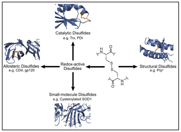
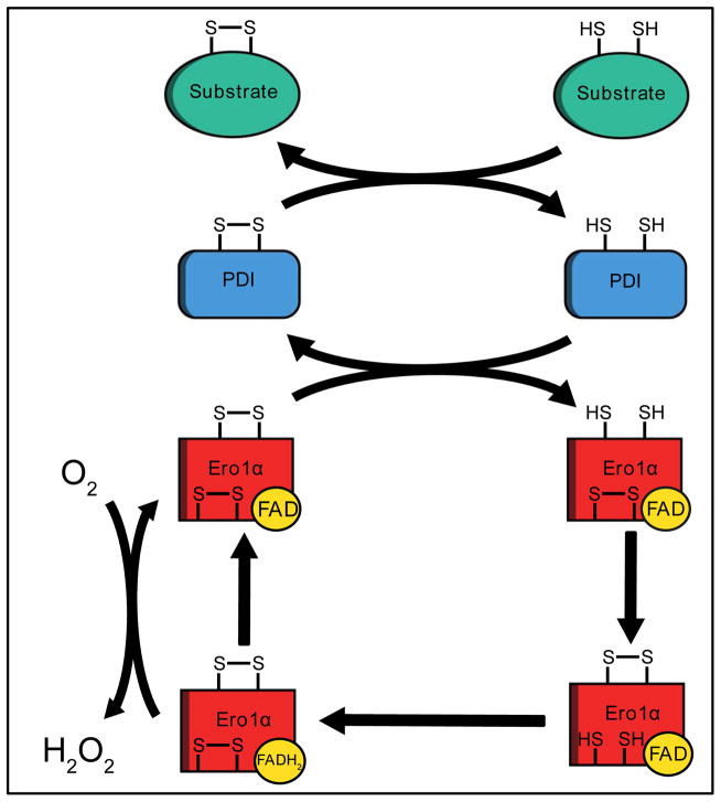
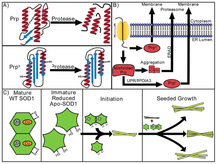
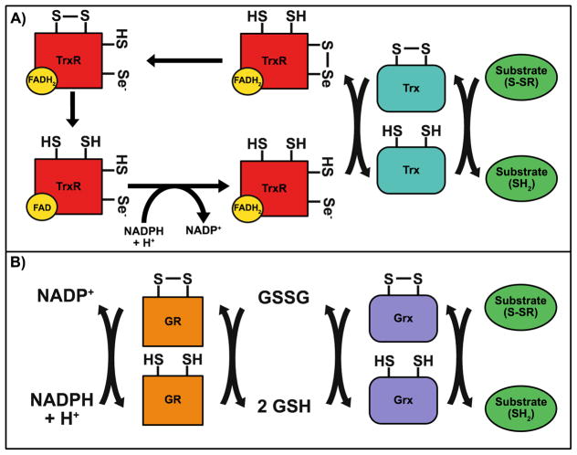
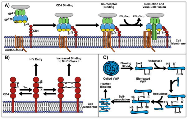
This review provides a comprehensive overview of the functional roles of disulfide bonds and their relevance to human disease. The critical roles of disulfide bonds in protein structure stabilization and redox regulation of protein activity are addressed. Disulfide bonds are essential to the structural stability of many proteins within the secretory pathway and can exist as intramolecular or inter-domain disulfides. The proper formation of these bonds often relies on folding chaperones and oxidases such as members of the protein disulfide isomerase (PDI) family. Many of the PDI family members catalyze disulfide-bond formation, reduction, and isomerization through redox-active disulfides and perturbed PDI activity is characteristic of carcinomas and neurodegenerative diseases. In addition to catalytic function in oxidoreductases, redox-active disulfides are also found on a diverse array of cellular proteins and act to regulate protein activity and localization in response to oxidative changes in the local environment. These redox-active disulfides are either dynamic intramolecular protein disulfides or mixed disulfides with small-molecule thiols generating glutathionylation and cysteinylation adducts. The oxidation and reduction of redox-active disulfides are mediated by cellular reactive oxygen species and activity of reductases, such as glutaredoxin and thioredoxin. Dysregulation of cellular redox conditions and resulting changes in mixed disulfide formation are directly linked to diseases such as cardiovascular disease and Parkinson's disease.


Figures

Figure 1
Figure 1
Characterized functional roles of disulfides: structural and redox-active disulfides. Redox-active disulfides may be further characterized as small-molecule, catalytic and allosteric disulfides.
Figure 2
Figure 2
The Ero1α-PDI-mediated oxidative protein-folding pathway. PDI is reoxidized by Ero1α, a FAD-binding oxidase. Ero1α transfers electrons from PDI to FAD with oxygen being the final electron acceptor.
Figure 3
Figure 3
Structural disulfides in disease. (A) The disulfide bond in Prpc is reduced in Prpsc, which is protease-resistant. (B) Misfolded Prp is removed from the ER and degraded via ERAD. Accumulation of misfolded Prp leads to activation of the UPR, resulting in increased expression of folding chaperones such as PDIA3, which interacts with misfolded Prp to re-fold and prevent aggregation. (C) Immature and disulfide-reduced apo-SOD1 or Zn-SOD1 initiates the formation of cross-β-amyloid fibrils creating a “nucleus.” The “nucleus” fragments to produce “seeds” that convert soluble SOD1 into insoluble β-rich structures, elongating the cross-β-amyloid fibrils.
Figure 4
Figure 4
The Trx-TrxR and Grx-GR-mediated disulfide-reduction pathways. (A) Upon reduction of substrate proteins, the resulting oxidized Trx is reduced by TrxR. TrxR transfers electrons from NADPH, via a bound FAD and a cysteine-cysteine and cysteine-selenocysteine couple to the oxidized Trx. (B) Similar to Trx, Grx reduces disulfide bonds in substrate proteins. Grx is reduced by GSH, yielding GSSG. GSH levels are restored by GR via the transfer of electrons from NADPH to GSSG via a redox-active disulfide on GR..
Figure 5
Figure 5
Redox-active disulfides in disease. (A) Reduction of a disulfide bond in gp120 by PDI or Trx promotes HIV virus-host cell fusion by conformational changes that expose the fusion peptide on gp41 for membrane insertion. (B) Reduction of a disulfide bond on CD4 results in the formation of CD4 homodimers linked by disulfide bonds. Dimeric CD4 shows increased binding to MHC Class II. The reduced monomer of CD4 is preferred for HIV entry. (C) The shear force of flowing blood induces conformational changes in VWF from a coiled ball to an elongated structure. Intramolecular disulfides in elongated VWF may be reduced by an oxidoreductase, promoting intermolecular disulfide bond formation and the self-association of VWF, which promotes platelet binding.
Figure 6
Figure 6
Proteomic methods to study redox-active disulfides. (A) The OxICAT method incorporates light and heavy ICAT reagents (inset) to reduced and oxidized cysteines, respectively, for analysis by MS. (B) isoTOP-ABPP utilizes a cysteine-reactive iodoacetamide-alkyne (IAA) probe (inset) to identify reactive cysteines within a proteome. Cysteine oxidation to form disulfides is accompanied by a characteristic loss in cysteine reactivity that can be quantified by incorporation of light and heavy tags (Azo-tags) to IAA-modified peptides for MS analysis. (C) Caged-bromomethylketone (Caged-BK) contains a masked cysteine-reactive electrophile that can be activated in situ by UV irradiation.
Figure 7
Figure 7
Proteomic methods to study glutathionylated proteins. (A) Glutathionylation results in the formation of a mixed disulfide between a protein and glutathione. (B) Biotinylated glutathione adducts used to study glutathionylation include BioGSH, BioGEE and N3GSH. (C) Glutathionylated proteins can be identified by capping reduced cysteines with NEM followed by Grx-mediated reduction of glutathionylated cysteines. The resulting newly-formed thiols are then capped with NEM-biotin for avidin-biotin enrichment of glutathionylated proteins.

References

    1. Sevier CS, Kaiser CA. Formation and transfer of disulphide bonds in living cells. Nat Rev Mol Cell Biol. 2002;3:836–847. - PubMed
    1. Feige MJ, Hendershot LM. Disulfide bonds in ER protein folding and homeostasis. Curr Opin Cell Biol. 2011;23:167–175. - PMC - PubMed
    1. Hatahet F, Ruddock LW. Protein disulfide isomerase: a critical evaluation of its function in disulfide bond formation. Antioxid Redox Signal. 2009;11:2807–2850. - PubMed
    1. Deponte M, Hell K. Disulphide bond formation in the intermembrane space of mitochondria. J Biochem. 2009;146:599–608. - PubMed
    1. Parakh S, Atkin JD. Novel roles for protein disulphide isomerase in disease states: a double edged sword? Front Cell Dev Biol. 2015;3:30. - PMC - PubMed

Publication types