. 2017 Jan 3:8:14128.
doi: 10.1038/ncomms14128.

Cleavage of DFNA5 by caspase-3 during apoptosis mediates progression to secondary necrotic/pyroptotic cell death

Affiliations

Cleavage of DFNA5 by caspase-3 during apoptosis mediates progression to secondary necrotic/pyroptotic cell death

Corey Rogers et al. Nat Commun. .

Abstract

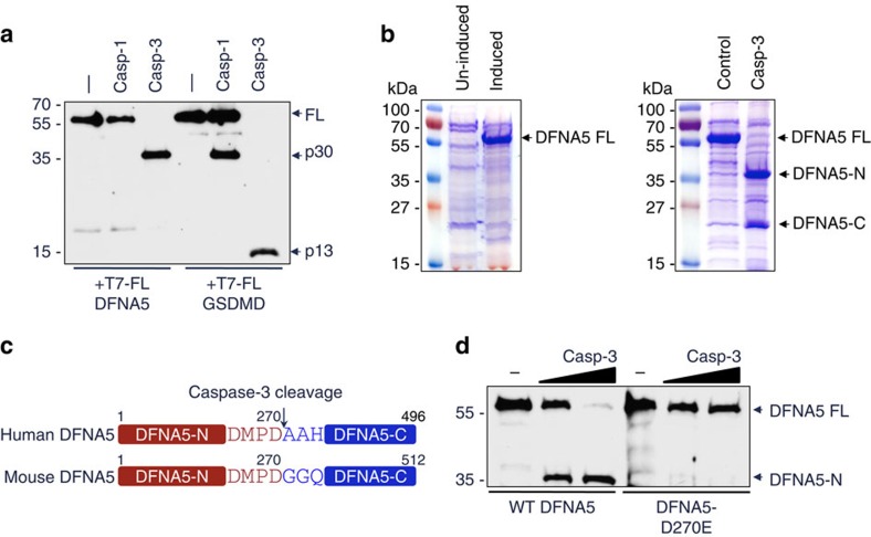
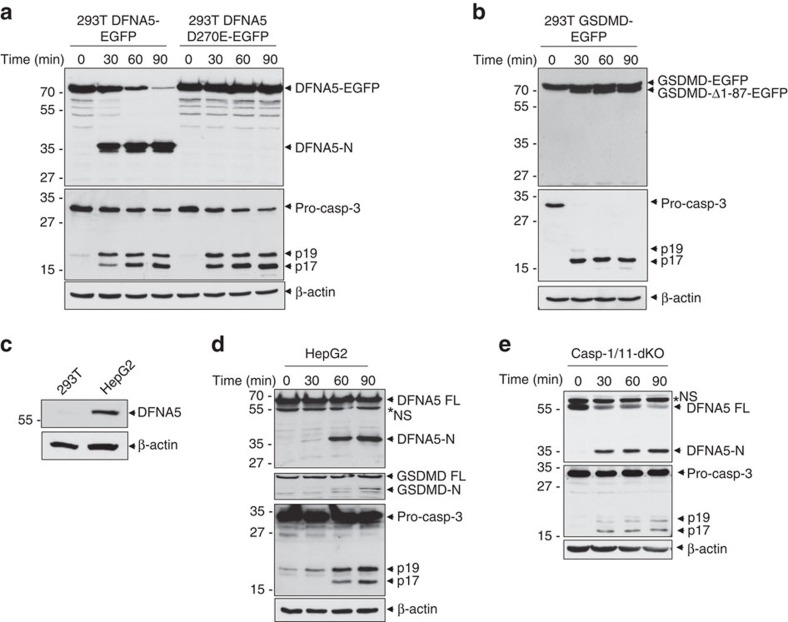
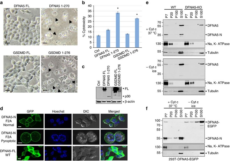
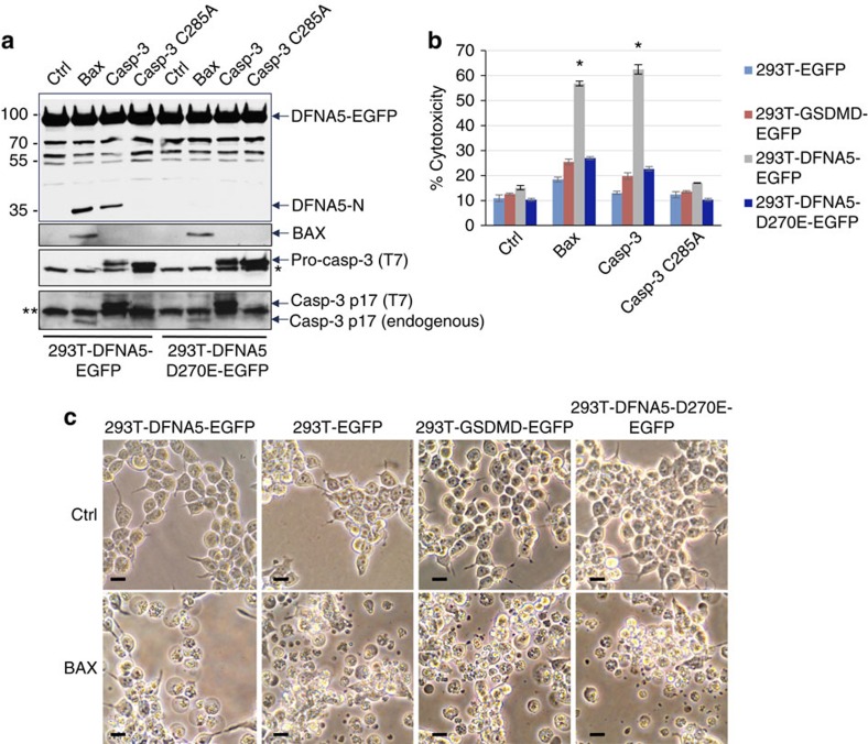
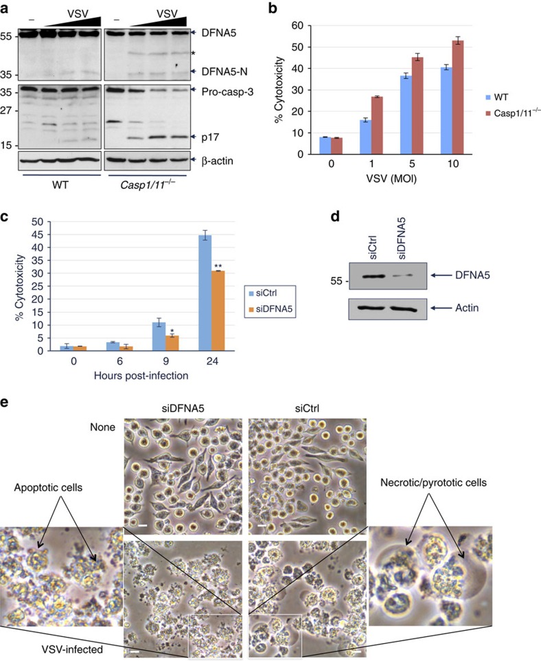
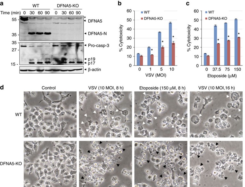
Apoptosis is a genetically regulated cell suicide programme mediated by activation of the effector caspases 3, 6 and 7. If apoptotic cells are not scavenged, they progress to a lytic and inflammatory phase called secondary necrosis. The mechanism by which this occurs is unknown. Here we show that caspase-3 cleaves the GSDMD-related protein DFNA5 after Asp270 to generate a necrotic DFNA5-N fragment that targets the plasma membrane to induce secondary necrosis/pyroptosis. Cells that express DFNA5 progress to secondary necrosis, when stimulated with apoptotic triggers such as etoposide or vesicular stomatitis virus infection, but disassemble into small apoptotic bodies when DFNA5 is deleted. Our findings identify DFNA5 as a central molecule that regulates apoptotic cell disassembly and progression to secondary necrosis, and provide a molecular mechanism for secondary necrosis. Because DFNA5-induced secondary necrosis and GSDMD-induced pyroptosis are dependent on caspase activation, we propose that they are forms of programmed necrosis.


Figures

Figure 1
Figure 1. Caspase-3 cleaves DFNA5 after Asp270.
(a) Immunoblot of purified N-terminal His6-T7-tagged DFNA5 and GSDMD proteins incubated without (−) or with caspase-1 (casp-1) or caspase-3 (casp-3) for 45 min at 37 °C. The blot was probed with anti-T7 antibody. (b) Coomassie stained SDS-polyacrylamide gels of TALON-immobilized proteins from uninduced or IPTG-induced BL-21 pET28b-DFNA5 bacteria (left panel), or IPTG-induced BL-21 pET28b-DFNA5 bacteria incubated without (control) or with caspase-3 for 45 min (right panel). The caspase-3-generated N- and C-terminal DFNA5 fragments (DFNA5-N and DFNA5-C, respectively) are indicated. (c) Diagrammatic representation of human and mouse DFNA5 proteins showing the caspase-3 recognition motif at aa 267–270. (d) Immunoblot of purified N-terminal T7-tagged WT DFNA5 and DFNA5-D270E proteins incubated without (−) or with increasing amounts of caspase-3 for 45 min. The blot was probed with anti-T7 antibody. Results are representative of at least three independent experiments.
Figure 2
Figure 2. DFNA5 is cleaved by caspase-3 downstream of the Apaf-1 apoptosome.
(a,b) Immunoblots of S100 lysates from stable 293T-DFNA5-EGFP and 293T-DFNA5-D270E-EGFP cells (a), or stable 293T-GSDMD-EGFP cells (b) stimulated with cytochrome c for the indicated times at 37 °C. The blots were probed with anti-DFNA5 (a, upper), anti-GSDMD (b, upper), anti-caspase-3 (a,b, middle) or anti-β-actin (a,b, lower) antibodies. (c) Immunoblot of endogenous DFNA5 in 293T and HEPG2 total cell lysates. (c,d) Immunoblots of S100 lysates from human HEPG2 (d) or mouse casp-1/casp-11-double knockout (casp-1/11-dKO) (e) macrophages stimulated with cytochrome c for the indicated times at 37 °C. The blots were probed with anti-DFNA5 (upper in d,e), anti-GSDMD (second from top in d), anti-caspase-3 (third from top in d, middle in e) or anti-β-actin (fourth from top in d, lower in e) antibodies. Asterisk indicate non-specific band (NS). Results are representative of at least three independent experiments.
Figure 3
Figure 3. The cleaved N-terminal DFNA5 fragment has intrinsic necrotic activity.
(a) Microscopic images of 293T cells transfected with expression constructs for full-length DFNA5 (DFNA5 FL), full-length GSDMD (GSDMD FL), DFNA5-N (DFNA5 1–270) or GSDMD-N (GSDMD 1–276) as indicated. Arrowheads indicate ballooned cell membrane characteristic of necrotic/pyroptotic cells. Scale bar, 20 μm. (b) Cytotoxicity of DFNA5 and GSDMD and their fragments as measured by LDH release in the culture supernatants of 293T cells transfected with the indicated expression constructs for DFNA5 and GSDMD. (n=3) *P<0.001, Student's t-test. (c) Immunoblots showing the expression of the proteins in (b). The lower expression of DFNA5 1–270 and GSDMD 1-276 fragments is due to their toxicity. (d) Confocal images of the DFNA5-N-F2A-EGFP mutant (upper and middle), or full-length WT DFNA5-EGFP expressed in 293T. The upper panels show normal cells and the middle panels show necrotic/pyroptotic cells. For videos of the cells shown in the middle panels see Supplementary Videos 1–3. Scale bar, 10 μm. (e,f) Immunoblots of cytochrome c-activated (+ Cyt c, 37° C) or unactivated (− Cyt c, ice) cell lysates from DFNA5+/+ (WT) and DFNA5−/− (DFNA5-KO) macrophages (e) or 293T-DFNA5-EGFP cells (f) probed with anti-DFNA5 (upper), anti-Na, K-ATPase (middle) or anti-tubulin (lower) antibodies after fractionation into P7 (heavy membrane), P20 (light membrane), P100 (insoluble cytosol) and S100 (soluble cytosol). Asterisk indicates non-specific band with similar size/migration as full-length DFNA5. Results are representative of at least three independent experiments. Error bars represent standard error of the mean (s.e.m).
Figure 4
Figure 4. Molecular determinants of plasma membrane targeting of DFNA5-N.
(a) Predicted structure of DFNA5 MTD spanning residues 1–57 showing the side chains of residues F2, K39, K40, K41, R42 and R48 as sticks. (b) Helical wheel representations of DNFA5 residues 1–14 showing the putative amphipathic nature of the N-terminal helix. K4, R11 and R7 are clustered on the left while F2 and F9 are on the right hand side of the helix. (cg) Confocal images of the indicated DFNA5-N-EGFP alanine mutants expressed in 293T. The ribbon diagrams on the left of each image show the position of side chains of residues mutated to alanines. Scale bar, 10 μm. (h) Cytotoxicity of the indicated DFNA5-N mutants as measured by LDH release in the culture supernatants of 293T cells transfected with constructs encoding these mutants (n=3). *P<0.001, Student's t-test. Expression of these mutants in cell lysates is shown in the top panel as measured by immunoblot analysis with anti-DFNA5 antibody. Results are representative of at least three independent experiments. Error bars represent standard error of the mean (s.e.m).
Figure 5
Figure 5. Activation of DFNA5 downstream of the mitochondrial apoptotic pathway.
(a) Immunoblots of cell lysates from stable 293T-DFNA5-EGFP and 293T-DFNA5-D270E-EGFP cells transfected with vector control (Ctrl) or constructs for Bax, constitutively active T7-tagged caspase-3 (casp-3) or inactive caspase-3 (casp-3-C285A) for 24 h as indicated. The blots were probed with anti-DFNA5 (upper), anti-Bax (middle), or anti-caspase-3 (lower two panels) antibodies. Asterisk indicates endogenous procaspase-3. Double asterisks indicate non-specific band. (b) Cytotoxicity of Bax and constitutively active caspase-3 as measured by LDH release in the culture supernatants of the indicated 293T cell lines. (n=3) *P<0.0001), Student's t-test. (c) Microscopic images of the indicated 293T cell lines transfected with a vector control (Ctrl) or an expression construct for full-length Bax (BAX). Scale bar, 20 μm. Results are representative of at least three independent experiments. Error bars represent standard error of the mean (s.e.m).
Figure 6
Figure 6. Activation of DFNA5 downstream of viral infection in macrophages.
(a) Immunoblots of DFNA5 in cell lysates from uninfected (−) or VSV-infected (1, 5, 10 MOI) WT (left) or casp-1/11 dKO (right) immortalized BMDMs. The lower panels show caspase-3 and β-actin in the same cell lysates. Asterisk indicates non-specific band. (b,c) Cytotoxicity of VSV as measured by LDH release in the culture supernatants of WT and casp-1/11 dKO BMDMs infected with VSV for 12 h (b) (n=3) or casp-1/11 dKO BMDMs transfected with control siRNA (siCtrl) or DFNA5 siRNA (siDFNA5) for 48 h and then infected with VSV (1 MOI) for the indicated times (c). (n=3) *P<0.01, **P<0.001, Student's t-test. (d) Immunoblots of DFNA5 in the control siRNA (siCtrl)- or DFNA5 siRNA (siDFNA5)-transfected casp-1/11 dKO BMDMs shown in (c). (e) Microscopic images of the control siRNA (siCtrl)- or DFNA5 siRNA (siDFNA5)-transfected casp-1/11 dKO BMDMs shown in (c). Scale bar, 20 μm. The insets show higher magnification images of apoptotic and necrotic cells. Results are representative of at least three independent experiments. Error bars represent standard error of the mean (s.e.m).
Figure 7
Figure 7. DFNA5 induces secondary necrosis downstream of viral infection in 293T cells.
(a,d) Cytotoxicity of VSV (a) (n=3) (*P<0.001; **P<0.0001, Student's t-test) or ECMV (d) (n=3) (*P<0.0001, Student's t-test) as measured by LDH release in the culture supernatants of the indicated 293T cell lines after 18 h infection with these viruses. (b,e) Immunoblots of DFNA5 in cell lysates from the indicated uninfected (−), VSV-infected (MOI: 1, 5 for 18 h) (b) or ECMV-infected (MOI: 0.005, 0.01, 0.05 for 18 h) (e) 293T cell lines. (c) Microscopic images of the indicated uninfected (Un) or VSV-infected (VSV) 293T cell lines. Scale bar, 20 μm. Results are representative of at least three independent experiments. Error bars represent standard error of the mean (s.e.m).
Figure 8
Figure 8. Knockout of DFNA5 reduces secondary necrosis in macrophages.
(a) Immunoblots of S100 lysates from DFNA5+/+ (WT) and DFNA5−/− (DFNA5-KO) macrophages stimulated with cytochrome c for the indicated times at 37 °C. The blots were probed with anti-DFNA5 (upper), anti-caspase-3 (middle) or anti-β-actin (lower) antibodies. Asterisk indicate non-specific band (NS). (b,c) Cytotoxicity of VSV (b) (n=3) and etoposide (c) (n=3) as measured by LDH release in the culture supernatants of DFNA5+/+ (WT) and DFNA5−/− (DFNA5-KO) macrophages infected with VSV or treated with etoposide for 8 h. *P<0.0001, Student's t-test. (d) Microscopic images of DFNA5+/+ (WT) and DFNA5−/− (DFNA5-KO) macrophages infected with VSV or treated with etoposide as indicated. White arrowheads indicate ballooning necrotic cells, and black arrowheads indicate blebbing apoptotic cells. Scale bar, 20 μm. Results are representative of at least three independent experiments. Error bars represent standard error of the mean (s.e.m).
Figure 9
Figure 9. Signalling pathways leading to activation of DFNA5.
Various death stimuli or viral infection can lead to permeabilization of the outer mitochondrial membrane causing the release of cytochrome c, which binds to Apaf-1 leading to assembly of the Apaf-1 apoptosome and activation of caspase-9. Within this complex active caspase-9 cleaves procaspase-3 to generate the active caspase-3 heterodimer. Active caspase-3 in turns cleaves DFNA5 at Asp270 to generate the necrotic DFNA5-N fragment which permeabilizes the plasma membrane by forming large pores causing osmotic lysis of the cell and releasing cellular contents including pro-inflammatory mediators and alamins, into the extracellular space. Caspase-3 can also be activated by the death receptor pathway, which is activated by death receptor ligands at the cell membrane. This pathway can potentially lead to caspase-3-mediated processing of DFNA5 and secondary necrosis.

Comment in

  • Secondary Necrosis: Accidental No More.
    Galluzzi L, Kroemer G. Galluzzi L, et al. Trends Cancer. 2017 Jan;3(1):1-2. doi: 10.1016/j.trecan.2016.12.001. Epub 2017 Jan 3. Trends Cancer. 2017. PMID: 28718422

References

    1. Wallach D., Kang T. B., Dillon C. P. & Green D. R. Programmed necrosis in inflammation: toward identification of the effector molecules. Science 352, aaf2154 (2016). - PubMed
    1. Linkermann A. & Green D. R. Necroptosis. N. Engl. J. Med. 370, 455–465 (2014). - PMC - PubMed
    1. Miao E. A., Rajan J. V. & Aderem A. Caspase-1-induced pyroptotic cell death. Immunol. Rev. 243, 206–214 (2011). - PMC - PubMed
    1. Shi J. et al.. Inflammatory caspases are innate immune receptors for intracellular LPS. Nature 514, 187–192 (2014). - PubMed
    1. Guo H., Callaway J. B. & Ting J. P. Inflammasomes: mechanism of action, role in disease, and therapeutics. Nat. Med. 21, 677–687 (2015). - PMC - PubMed

Publication types

MeSH terms