Review
. 2017 Jan 3;127(1):74-82.
doi: 10.1172/JCI88883. Epub 2017 Jan 3.

The ominous triad of adipose tissue dysfunction: inflammation, fibrosis, and impaired angiogenesis

Review

The ominous triad of adipose tissue dysfunction: inflammation, fibrosis, and impaired angiogenesis

Clair Crewe et al. J Clin Invest. .

Abstract

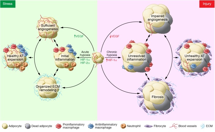
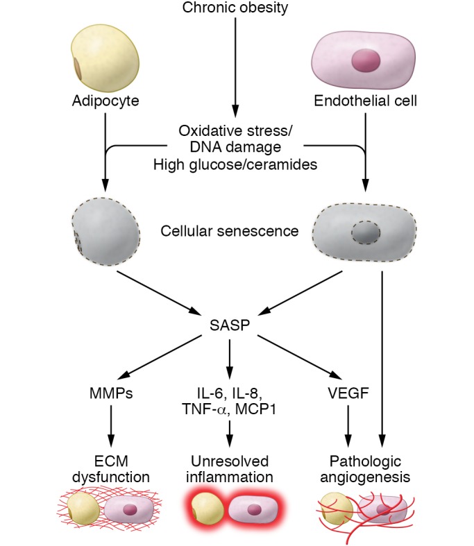
There are three dominant contributors to the pathogenesis of dysfunctional adipose tissue (AT) in obesity: unresolved inflammation, inappropriate extracellular matrix (ECM) remodeling and insufficient angiogenic potential. The interactions of these processes during AT expansion reflect both a linear progression as well as feed-forward mechanisms. For example, both inflammation and inadequate angiogenic remodeling can drive fibrosis, which can in turn promote migration of immune cells into adipose depots and impede further angiogenesis. Therefore, the relationship between the members of this triad is complex but important for our understanding of the pathogenesis of obesity. Here we untangle some of these intricacies to highlight the contributions of inflammation, angiogenesis, and the ECM to both "healthy" and "unhealthy" AT expansion.


Conflict of interest statement

The authors have declared that no conflict of interest exists.

Figures

Figure 1
Figure 1. The complex contributions of angiogenesis, inflammation, and fibrosis in healthy and unhealthy AT expansion.
Hypoxia during early AT expansion induces stress signaling that facilitates angiogenesis via upregulation of an array of genes including VEGF, initiates inflammatory cell infiltration, and induces organized ECM remodeling to encourage appropriate AT expansion. However, persistent stress signaling translates into injury signaling, caused by long-term and severe hypoxia. This hypoxic condition fails to induce VEGF, resulting in impaired angiogenesis. Limited oxygen supply also triggers chronic low-grade inflammation predominantly characterized by proinflammatory macrophage infiltration. Upregulation of HIF-1α also increases the fibrotic program in AT. The integrated responses among those three factors promote significant hypotrophy in adipose tissue, and the consequence is an unhealthy AT expansion as well as obesity-associated metabolic consequences.
Figure 2
Figure 2. The role of senescence in obesity-associated AT dysfunction.
Chronic obesity can cause AT oxidative stress, DNA damage, and increased exposure to high glucose and ceramide concentrations. These deleterious factors can drive cellular senescence in many cells types. Adipocyte and endothelial cell senescence have been specifically studied in the context of obesity. Mature adipocytes and endothelial cells as well as their precursors can undergo senescence and take on the SASP. SASP factors can promote AT dysfunction through dysregulation of AT ECM remodeling, inflammation, and angiogenesis.

References

    1. Li J, Yu X, Pan W, Unger RH. Gene expression profile of rat adipose tissue at the onset of high-fat-diet obesity. Am J Physiol Endocrinol Metab. 2002;282(6):E1334–E1341. doi: 10.1152/ajpendo.00516.2001. - DOI - PubMed
    1. Kleemann R, et al. Time-resolved and tissue-specific systems analysis of the pathogenesis of insulin resistance. PLoS One. 2010;5(1):e8817. doi: 10.1371/journal.pone.0008817. - DOI - PMC - PubMed
    1. Lee YS, et al. Increased adipocyte O2 consumption triggers HIF-1α, causing inflammation and insulin resistance in obesity. Cell. 2014;157(6):1339–1352. doi: 10.1016/j.cell.2014.05.012. - DOI - PMC - PubMed
    1. Halberg N, et al. Hypoxia-inducible factor 1α induces fibrosis and insulin resistance in white adipose tissue. Mol Cell Biol. 2009;29(16):4467–4483. doi: 10.1128/MCB.00192-09. - DOI - PMC - PubMed
    1. Appelhoff RJ, et al. Differential function of the prolyl hydroxylases PHD1, PHD2, and PHD3 in the regulation of hypoxia-inducible factor. J Biol Chem. 2004;279(37):38458–38465. doi: 10.1074/jbc.M406026200. - DOI - PubMed

Publication types