. 2017 Apr;11(4):972-987.
doi: 10.1038/ismej.2016.169. Epub 2017 Jan 3.

The Pseudomonas putida T6SS is a plant warden against phytopathogens

Affiliations

The Pseudomonas putida T6SS is a plant warden against phytopathogens

Patricia Bernal et al. ISME J. 2017 Apr.

Abstract

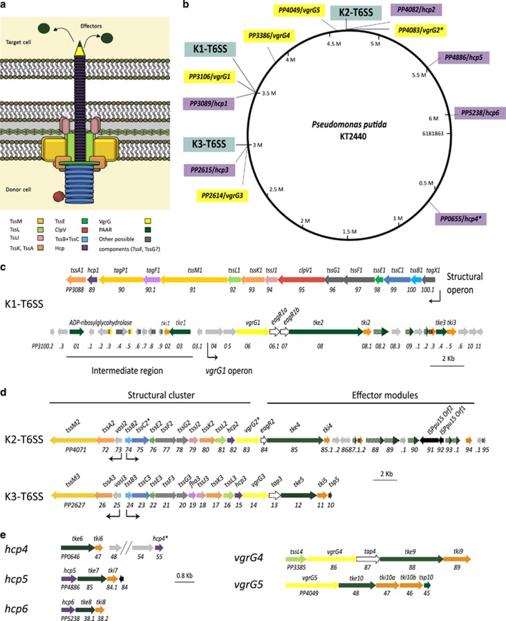
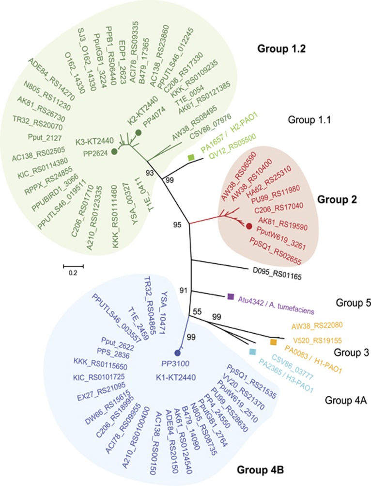
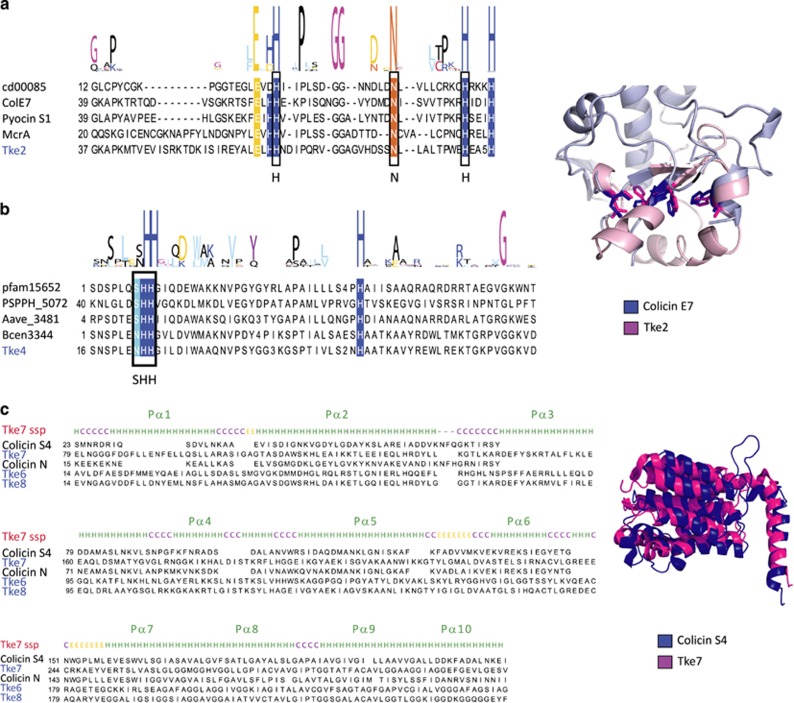
Bacterial type VI secretion systems (T6SSs) are molecular weapons designed to deliver toxic effectors into prey cells. These nanomachines have an important role in inter-bacterial competition and provide advantages to T6SS active strains in polymicrobial environments. Here we analyze the genome of the biocontrol agent Pseudomonas putida KT2440 and identify three T6SS gene clusters (K1-, K2- and K3-T6SS). Besides, 10 T6SS effector-immunity pairs were found, including putative nucleases and pore-forming colicins. We show that the K1-T6SS is a potent antibacterial device, which secretes a toxic Rhs-type effector Tke2. Remarkably, P. putida eradicates a broad range of bacteria in a K1-T6SS-dependent manner, including resilient phytopathogens, which demonstrates that the T6SS is instrumental to empower P. putida to fight against competitors. Furthermore, we observed a drastically reduced necrosis on the leaves of Nicotiana benthamiana during co-infection with P. putida and Xanthomonas campestris. Such protection is dependent on the activity of the P. putida T6SS. Many routes have been explored to develop biocontrol agents capable of manipulating the microbial composition of the rhizosphere and phyllosphere. Here we unveil a novel mechanism for plant biocontrol, which needs to be considered for the selection of plant wardens whose mission is to prevent phytopathogen infections.


Conflict of interest statement

The authors declare no conflict of interest.

Figures

Figure 1
Figure 1
T6SS clusters in P. putida KT2440. (a) Schematic representation of the T6SS structure. (b) Distribution of the K1-, K2- and K3-T6SS clusters (blue), and the vgrG (yellow) and hcp (purple) genes in the KT2440 genome. (ce) Genomic organization of the P. putida T6SSs cluster, including K1 (c), K2 and K3 (d) or the vgrG and hcp orphan clusters (e). The color code of the genes correlates with the color code shown in a. The asterisk (*) in the tssC2, vgrG2 and hcp4 genes indicates that these genes contain premature stop codons.
Figure 2
Figure 2
Phylogenetic distribution of T6SS clusters in P. putida species. Maximum likelihood tree with 1000 bootstrap replicates were built with Mega 6 for the core component protein TssB. T6SS cluster nomenclature (Boyer et al., 2009; Barret et al., 2011) is used to show the major phylogenetic clusters. Three main groups are clearly distinguishable: group 1.2 (green), group 2 (red) and group 4B (blue). P. aeruginosa and A. tumefaciens T6SSs loci are included into the phylogenetic tree to illustrate all the subgroups: 1.1 (P. aeruginosa H2), 1.2 (P. putida K2-K3), 2 (P. putida W619), 3 (P. aeruginosa H1), 4A (P. aeruginosa H3), 4B (P. putida K1) and 5 (A. tumefacines).
Figure 3
Figure 3
Functionality of the P. putida K1-T6SS. (a) Production and secretion of Hcp1 in the P. putida KT2440 wild type and the ΔtssA1 mutant strains. The HA-tagged Hcp1 protein was detected by western blot analysis using an anti-HA antibody. Detection of the β-subunit of the RNA polymerase (β-RNAP) was used as control. The position of the molecular size marker (in kDa) is indicated. (b) Competition assay between P. putida and a lacZ-encoding E. coli strain. Blue patches on X-gal-containing LB plates indicate E. coli survival. The top row shows the growth of E. coli, P. putida KT2440 wild-type strain and a battery of P. putida mutants in K1-T6SS genes. The bottom row shows the growth of mixed E. coli/P. putida cultures after 5 h of co-incubation.
Figure 4
Figure 4
P. putida KT2440 T6SS effectors. (a) The domain organization of the putative effectors is shown, with PAAR motifs indicated in orange, MIX motifs in blue, Rhs domains in green, HNH nuclease motifs (Tox-HNH and Tox-SHH) in purple, colicin motifs in yellow and the Tox-61 domain in pink. Multiple sequence alignments of the PAAR (b) and MIX (c) protein motifs are represented. The KT2440 T6SS effectors identified in this work are indicated in blue. The sequence of known T6SS effectors containing these motifs was retrieved from the NCBI database (http://www.ncbi.nlm.nih.gov/Structure/cdd/cdd.shtml). Conservation logos of the motifs are indicated above the alignment. Conserved residues are highlighted according to the amino acid characteristic: hydrophobic (black), small (pink), positive (blue), negative (yellow) and polar (purple, light blue, red).
Figure 5
Figure 5
P. putida KT2440 T6SS nucleases. (a, b) Multiple sequence alignments of the C-terminal domains of Tke2 (a) and Tke4 (b) effectors (blue) with known proteins of the family (black). Conservation logos of the motifs HNH (a) and SHH (b) are indicated above the alignment. Conserved residues are indicated with the color code used in Figure 4. A representation of the structural model of the C-terminal domain of the Tke2 effector (magenta) superimposed on the colicin E7 structure (blue; PDB: 2JB0) is shown on the right of a. Side chains of the active site residues are shown. (c) Multiple sequence alignment of T6SS colicin effectors (blue) with known proteins of the family (black). The secondary structure prediction (ssp) for effector Tke7 is shown above the alignment. A structural alignment of the Tke7 effector model (magenta) with the colicin S4 (blue, PDB: 3FEW) is shown on the right.
Figure 6
Figure 6
Toxicity and secretion of the Tke2 effector. (a) The growth of E. coli K12 cells harboring the pTke2-CT and pTki2 plasmids containing the C-terminal toxin domain of the tke2 effector and the tki2 immunity genes, respectively, was determined by measuring the OD at 600 nm. At time zero, either 1 mM isopropyl β-D-1-thiogalactopyranoside (IPTG) and/or 0.02% (w/v) arabinose were added to the LB medium, to induce expression of the tke2-CT or/and tki2 genes, respectively. (b) Western blot analyses using an anti-V5 or anti-HA monoclonal antibody to detect the Tke2-CT-V5 or Tki2-HA-tagged proteins. Proteins were prepared from E. coli K12 cells grown during 10 h in presence (+) or absence (−) of 1 mM IPTG and/or 0.02% (w/v) arabinose. (c) The indicated P. putida KT2440 strains bearing a tke2-V5-tagged gene were grown in tryptone soya broth (TSB) medium for 5 h. Tke2-V5 was detected in the whole cell and supernatant fractions using a monoclonal anti-V5 antibody. Detection of the β-subunit of the RNA polymerase (β-RNAP) was used as control. The position of the molecular size marker (in kDa) is indicated.
Figure 7
Figure 7
Bactericidal activity of P. putida KT2440 against a panel of phytopathogens. X. campestris, A. tumefaciens, P. carotovorum and P. syringae pv. tomato strains harbor the pRL662-gfp plasmid that confers gentamycin resistance. The P. putida KT2440 wild type (WT) and its isogenic ΔtssA1ΔtssM2ΔtssM3 triple mutant (ΔT6SS) were co-incubated with the phytopathogens for 24 h. Colony-forming unit (CFU) quantifications were performed on gentamycin selection. The average±s.d. from at least three biological replicates is plotted.
Figure 8
Figure 8
In planta competition assay between the biocontrol strain P. putida KT2440 and the phytopathogen X. campestris. (a) Leaves of N. benthamiana 24 h (upper panel) and 5 days (lower panel) after being infiltrated with X. campestris (pRL662-gfp; expressing a plasmid-encoded green fluorescence protein), the P. putida KT2440 wild type (WT), or its isogenic ΔtssA1ΔtssM2ΔtssM3 triple mutant (ΔT6SS). (b) Leaves of N. benthamiana 24 h (upper panel) and 5 days (lower panel) after co-infiltration of X. campestris (pRL662-gfp) with the indicated P. putida strain. In upper panel a and b, the leaves were visualized by fluorescence microscopy using a Leica M205FA stereomicroscope. The necrotic areas resulting from X. campestris infection are marked. The deep brown zone of necrosis is spread on a large portion of the leave (right panel), while such spread is far more restricted when the phytopathogen is co-inoculated with a T6SS positive P. putida strain (left panel). (c) Quantification of X. campestris (pRL662-gfp) colony-forming units (CFUs) recovered from N. benthamiana leaves after 24 h of co-infiltration with the indicated P. putida strain. X. campestris CFU were quantified after gentamycin (Gm) selection. Graphs represent mean +s.d., of at least five biological replicates with two technical replicates per experiment, statistical significance is indicated t-test P<0.001.

References

    1. Alcoforado Diniz J, Coulthurst SJ. (2015). Intraspecies competition in serratia marcescens is mediated by type VI-secreted Rhs effectors and a conserved effector-associated accessory protein. J Bacteriol 197: 2350–2360. - PMC - PubMed
    1. Alcoforado Diniz J, Liu Y-CC, Coulthurst SJ. (2015). Molecular weaponry: diverse effectors delivered by the Type VI secretion system. Cell Microbiol 17: 1742–1751. - PMC - PubMed
    1. Amer GA, Utkhede RS. (2000). Development of formulations of biological agents for management of root rot of lettuce and cucumber. Can J Microbiol 46: 809–816. - PubMed
    1. Aschtgen M-SS, Bernard CS, De Bentzmann S, Lloubès R, Cascales E. (2008). SciN is an outer membrane lipoprotein required for type VI secretion in enteroaggregative Escherichia coli. J Bacteriol 190: 7523–7531. - PMC - PubMed
    1. Aschtgen M-SS, Thomas MS, Cascales E. (2010). Anchoring the type VI secretion system to the peptidoglycan: TssL, TagL, TagP... what else? Virulence 1: 535–540. - PubMed

MeSH terms

Substances