. 2017 Jan 3;7(1):e991.
doi: 10.1038/tp.2016.254.

Hippocampal encoding of interoceptive context during fear conditioning

Affiliations

Hippocampal encoding of interoceptive context during fear conditioning

S-W Yoo et al. Transl Psychiatry. .

Abstract

Rodent models of auditory fear conditioning are often used to understand the molecular mechanisms regulating fear- and anxiety-related behaviors. Conditioning and extinction memories are influenced by contextual cues, and the reinstatement of conditioned fear occurs when the conditioning stimulus is presented in a context different from the extinction context. Although it has been proposed that internal state is a feature of context that could influence extinction, contributions of interoception to conditioning have not been experimentally addressed. Here we use ethanol (EtOH) to show that interoceptive cues are encoded through the hippocampus by mechanisms that involve increased phosphorylation of GluR1 on serine 845, and biophysical alterations in neuronal membranes that facilitate stabilization of surface-located calcium-permeable n-2-amino-3-(5-methyl-3-oxo-1,2-oxazol-4-yl) propanoic acid (AMPA) receptor (AMPAR) into membrane microdomains. Conflicting interoceptive cues during extinction and fear relapse testing resulted in a failure to consolidate extinction that was reversed by the administration of AMPAR antagonists immediately following the retrieval cue.


Conflict of interest statement

The authors declare no conflict of interest.

Figures

Figure 1
Figure 1
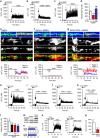
EtOH interferes with the consolidation of behavioral extinction. (a) Schematic illustration of the fear conditioning paradigm. (b) Freezing (% time) for each of the CS–US pairing trails during day 0 of fear conditioning, demonstrating an establishment of the CS–US pairing. (c) Freezing during the first and last 30 s of the retrieval period showing that EtOH did not interfere with the reconsolidation of fear conditioning. (d) Repeated CS exposure during extinction trials resulted in a gradual decrease in the CR that was similar in mice previously exposed to EtOH or water. (e) Fear relapse trials showing reinstatement of the CR in mice exposed to EtOH. Data are presented as mean±s.e.m. of n=20 per condition; significant differences between first 30 s and last 30 s in retrieval; between water and EtOH were determined by one-way ANOVA. *P<0.05, **P<0.01 compared with water. ANOVA, analysis of variance; CR, conditioned response; CS, conditioned stimulus; EtOH, ethanol; FC, fear conditioning; US, unconditioned stimulus.
Figure 2
Figure 2
EtOH enhances phosphorylation of GluR1 at serine 845 in hippocampus. Representative immunoblots of GluR1, GluR1S845, GluR1S831 and quantitative analysis of band density from HIP and LA following the indicated treatment conditions. (a, d) Twenty-four hours after FC (lanes indicated+are fear conditioned, and lanes indicated− are sham control). (b,e) Twenty-four hours after FC including 2 h of exposure to binge EtOH intake (ETOH) or water. (c,f) Twenty-four hours after FC including 2 h of exposure to binge EtOH intake or water, and following the retrieval cue (Ret; tone). (g,i) Immediately following extinction for the indicated treatment groups. (h, j) Twenty-four hours following extinction and immediately following the fear relapse test. Representative traces show relative changes in protein expression of GluR1, GluR1S831 and GluR1S845 for the indicated treatment conditions in HIP (k) and LA (l). Data are mean±s.d. *P<0.05, **P<0.01 compared with the indicated treatment condition. EtOH, ethanol; FC, fear conditioning; HIP, hippocampus; LA, lateral amygdala.
Figure 3
Figure 3
EtOH modifies the biophysical properties of cellular membranes and redistributes GluR1 to microdomains. (a) Representative immunofluorescent images showing EtOH facilitated a rapid (2 min) redistribution of the microdomains (GM1+, red) into enlarged clusters along dendrites, simultaneously resulted in redistributions of GluR1 (green) with clusters located to GM1+ microdomains (arrowheads indicate GM1/GluR1-co-localized microdomains). (b) Size of individual GM1+ microdomains, and (c) number of GluR1 located to GM1+ microdomains (n=276–440 microdomains from a minimum of 21 dendrites and 3 separate cultures per condition). (d) Representative immunoblots showing detergent-resistant membrane microdomains isolated by density centrifugation containing a membrane microdomain-enriched protein Flotillin (largely located to the more buoyant lipid-rich fractions, 1–3), a non-microdomain protein Transferrin (largely located outside of membrane microdomains, fractions 7–9) and GluR1. EtOH (2 min) induced a redistribution of GluR1 to membrane microdomains. (e) Representative immunoblots of surface biotin protein labeling and immunoprecipitation with subsequent immunoblotting for GluR1 or GluR2. (f) Quantitative analysis of immunoblots showing that EtOH specifically increased surface GluR1 but not total GluR1. Data are presented as mean±s.d. EtOH, ethanol. *P<0.05, ***P<0.001 compared with vehicle; #P<0.05, ###P<0.001 compared with EtOH. Scale bar, 20 μm.
Figure 4
Figure 4
Sensitization of focal AMPA-evoked calcium responses in membrane microdomains. AMPA-evoked calcium transients were measured along dendritic branches using the ratiometric calcium probe Fura-2 at the rate of 10 image pairs per second. Focal calcium bursts evoked by (a) AMPA (20 μM) were (b) suppressed when EtOH remained present in the bathing media, and (c) enhanced during EtOH WD. (d) Quantitation of AMPA-evoked calcium transients showing the median amplitudes of calcium responses for the indicated conditions. (e–g) Representative images for the indicated conditions showing (from top to bottom) pseudocolor images of baseline and AMPA-evoked calcium transients, immunofluorescent staining of the same dendrite for GM1, GluR1 and the merged images. Lower tracings show baseline and AMPA-evoked calcium responses for the indicated regions. (h–k) Representative traces of AMPA-evoked calcium transients evoked after a 2 min pre-exposure to vehicle or EtOH were inhibited by the selective calcium-permeable AMPAR antagonist, Naspm trihydrochloride (Naspm, 50 μM). (l) Quantification of AMPA-evoked calcium transients showing the median amplitudes of calcium currents evoked under the indicated conditions. (m) Representative immunoblots of rat hippocampal neurons treated with forskolin (Forsk, 20 μM) and IBMX (50 μM). (n) Quantification of surface GluR1 and total GluR1 after treatment of forskolin and IBMX. (o) Representative traces of AMPA-evoked calcium responses in neurons pretreated with forskolin and IBMX and then continuously exposed to EtOH. (p) Quantification of AMPA-evoked calcium responses for the indicated conditions. Data are median±s.d. ***P<0.001; ##P<0.01; ###P<0.001. AMPA, n-2-amino-3-(5-methyl-3-oxo-1,2-oxazol-4-yl) propanoic acid; AMPAR, AMPA receptor; EtOH, ethanol; IBMX, 3-isobutyl-1-methylxanthine; WD, withdrawal.
Figure 5
Figure 5
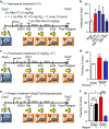
AMPAR antagonists rescue EtOH-associated impairment in the consolidation of extinction. (a) Schematic illustration of therapeutic time windows for Talampanel administration. Talampanel was intraperitoneally injected immediately following fear conditioning (i, 5 mg kg−1), three times over 18 h (ii, 6 h interval, 5 mg kg−1), or a single dose after retrieval (iii, 5 mg kg−1). Blue arrows depict dosing time and intervals. (b) Quantitative data showing freezing (% time) during fear relapse testing for the indicated treatment conditions. (c) Schematic illustration of timing for Peramapanel treatment. A single dose of Perampanel was intraperitoneally injected after retrieval (blue arrow, 5 mg kg−1). (d) Quantitative analysis of freezing behavior (% time) for the indicated treatment conditions. (e) Schematic illustration of timing for Naspm treatment. Mice received bilateral injections of Naspm into hippocampi (4 μg per 0.5 μl per min) after exposure to the retrieval cue. (f) Quantitative analysis of freezing behavior (% time) for the indicated treatment conditions. Data are mean±s.d. of n=10–14 animals per condition. *P<0.05, **P<0.01, #P<0.05 and ###P<0.001. ANOVA with Tukey post hoc comparisons. ANOVA, analysis of variance; AMPA, n-2-amino-3-(5-methyl-3-oxo-1,2-oxazol-4-yl) propanoic acid; AMPAR, AMPA receptor; EtOH, ethanol; FC, fear conditioning.

Similar articles

Cited by

References

    1. Lee JL. Memory reconsolidation mediates the strengthening of memories by additional learning. Nat Neurosci 2008; 11: 1264–1266. - PubMed
    1. Maren S. Seeking a spotless mind: extinction, deconsolidation, and erasure of fear memory. Neuron 2011; 70: 830–845. - PMC - PubMed
    1. Maren S, Phan KL, Liberzon I. The contextual brain:implications for fear conditioning, extinction and psychopathology. Nat Rev Neurosci 2013; 14: 417–428. - PMC - PubMed
    1. Johansen JP, Cain CK, Ostroff LE, LeDoux JE. Molecular mechanisms of fear learning and memory. Cell 2011; 147: 509–524. - PMC - PubMed
    1. Lee S, Song B, Kim J, Park K, Hong I, An B et al. GluA1 phosphorylation at serine 831 in the lateral amygdala is required for fear renewal. Nat Neurosci 2013; 16: 1436–1444. - PubMed

MeSH terms

LinkOut - more resources