. 2017 Jan 3;12(1):e0169081.
doi: 10.1371/journal.pone.0169081. eCollection 2017.

Induction of Multiple miR-200/182 Members in the Brains of Mice Are Associated with Acute Herpes Simplex Virus 1 Encephalitis

Affiliations

Induction of Multiple miR-200/182 Members in the Brains of Mice Are Associated with Acute Herpes Simplex Virus 1 Encephalitis

Anna Majer et al. PLoS One. .

Erratum in

Abstract

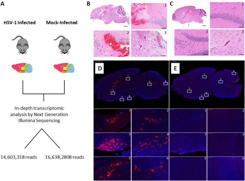
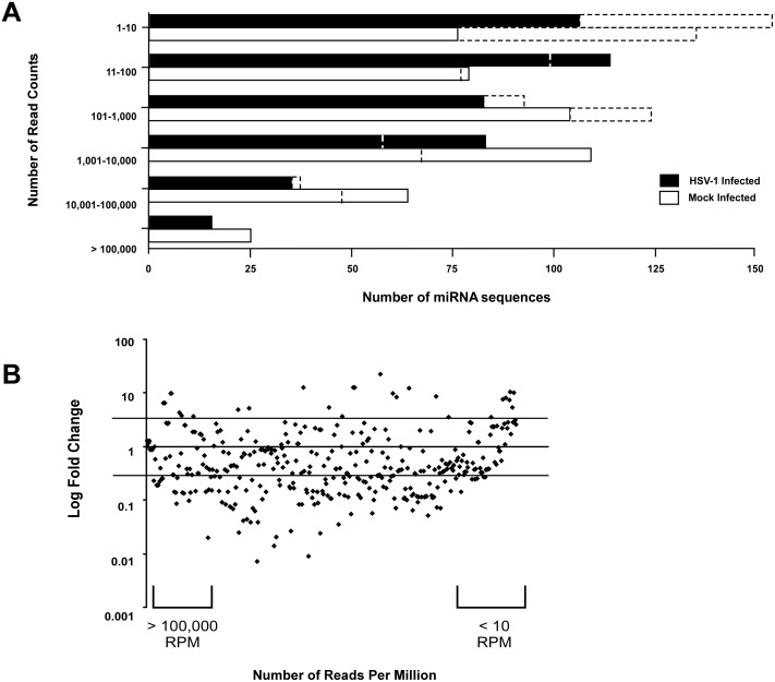
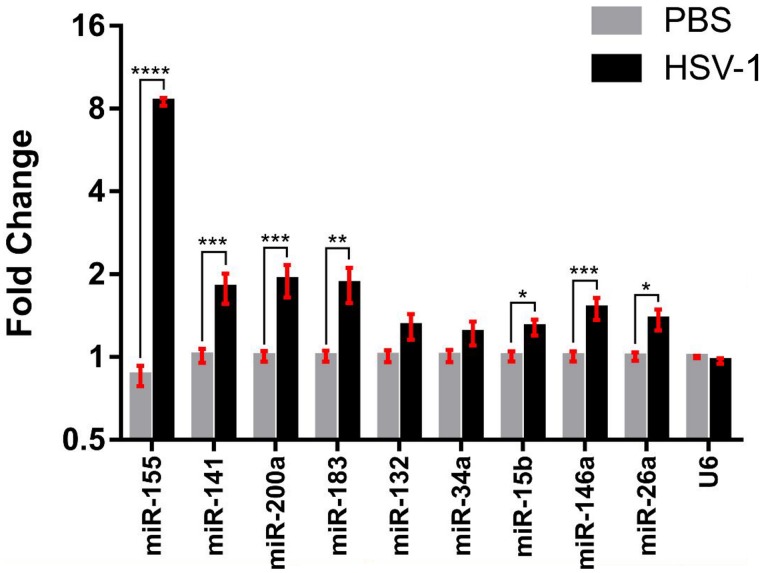
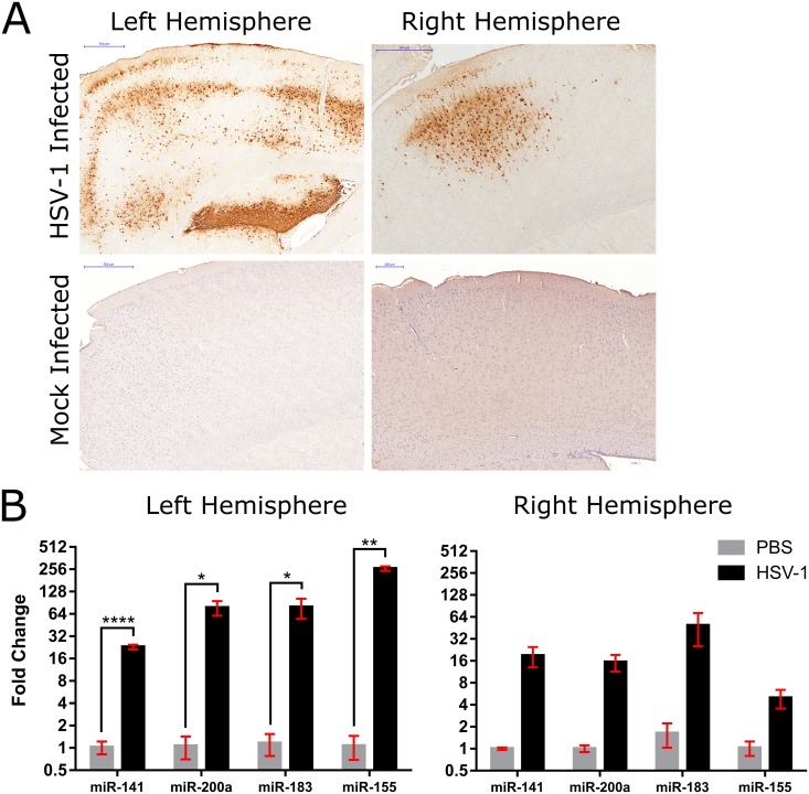
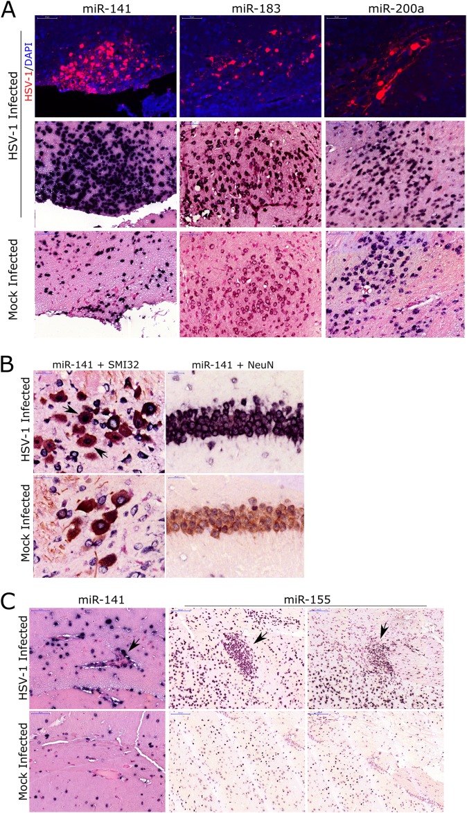
Important roles of microRNAs (miRNAs) in regulating the host response during viral infection have begun to be defined. However, little is known about the functional roles of miRNAs within an in vivo acute viral encephalitis model. We therefore identified global changes in miRNA expression during acute herpes simplex virus type 1 (HSV-1) encephalitis (HSVE) in mice. We found that many of the highly upregulated miRNAs (miR-155, miR-146a and miR-15b) detected in HSV-1 infected brain tissue are known regulators of inflammation and innate immunity. We also observed upregulation of 7 members belonging to the related group of miRNAs, the miR-200 family and miR-182 cluster (miR-200/182). Using in situ hybridization, we found that these miRNAs co-localized to regions of the brain with severe HSVE-related pathology and were upregulated in various cell types including neurons. Induction was apparent but not limited to cells in which HSV-1 was detected by immunohistochemistry, suggesting possible roles of these miRNAs in the host response to viral-induced tissue damage. Bioinformatic prediction combined with gene expression profiling revealed that the induced miR-200/182 members could regulate the biosynthesis of heparan sulfate proteoglycans. Using luciferase assays, we found that miR-96, miR-141, miR-183 and miR-200c all potentially targeted the syndecan-2 gene (Sdc2), which codes for a cell surface heparan sulfate proteoglycan involved in HSV-1 cellular attachment and entry.


Conflict of interest statement

The authors have declared that no competing interests exist.

Figures

Fig 1
Fig 1. Pathology in a mouse model of acute HSVE.
(A) Schematic representation of the animal experiments and process used in these studies to profile miRNAs by next generation sequencing. (B-C) Hematoxylin and Eosin staining depicting severe hemorrhaging and mononuclear cell infiltration within the blood vessels with prominent perivascular cuffing in (B) HSV-1 infected tissue. This pathogloy was not observed in (C) mock-infected mice inoculated with Vero cell suspension. Scale bars: low magnification images = 1000 μm; high magnification images = 50 μm. (D-E) Immunofluorescent images depicting HSV-1 positive cells (red) counterstained with DAPI (blue) in (D) HSV-1 and (E) mock-infected brain tissue. The magnified brain regions: (1) dentate gyrase, (2) caudate putamen, (3 and 4) hypothalamus regions, (5) pons and (6) cerebellum. Scale bars: low magnification images = 1000 μm; high magnification images = 50 μm.
Fig 2
Fig 2. Deregulation of miRNAs during HSVE.
(A) Number of miRNAs grouped based on number of read counts sequenced in HSV-1 and mock-infected samples, before normalization (dashed bars) and after normalization (solid bars) of sequencing data. (B) Mapping the fold change over the number of reads per million for each respective miRNA identified 50 upregulated and 166 downregulated miRNAs that were changed by equal to or more than 2-fold. (After filtering low abundance transcripts with less than 200 reads 24 miRNAs were upregulated and 54 downregulated) (C) The expression of HSV-1 encoded hsv1-miR-H1 was determined by the TaqMan miRNA assay in HSV-1 and mock-infected samples.
Fig 3
Fig 3. miR-200/182 and several others were upregulated in HSVE brain samples.
Fold change of miRNAs compared between HSV-1 infected and PBS treated whole brain tissues. U6 served as the normalization control for all validations. Data is represented as mean ± SEM (n = 4). A one-tailed unpaired t-test was used to calculate significance where * p<0.05; ** p<0.01; *** p<0.005; **** p<0.001.
Fig 4
Fig 4. Upregulation of miR-200/182 members and miR-155 was detected in HSV-1 infected cortex region.
(A) Immunohistochemistry images depicting the region of the cortex that was removed by laser capture microdissection from both left and right brain hemispheres of HSV-1 and mock-infected samples. HSV-1 positive cells are brown. Scale bar = 500 or 200 μm. (B) Fold change of miRNAs compared between HSV-1 infected and PBS treated cortical brain regions. Data is represented as mean ± SEM. A one-tailed unpaired t-test was used to calculate significance where * p<0.05; ** p<0.01; **** p<0.001.
Fig 5
Fig 5. Detection of miR-200/182 upregulation in HSVE brain tissues by in situ hybridization.
(A) Immunofluorescence images for HSV-1 (red) in HSV-1 and mock-infected brain tissue in relation to in situ hybridization for miR-141, miR-183 and miR-200a within similar brain areas. Hypothalamus region coinsides with miR-141 and miR-182 staining while pons region coinsides with staining for miR-200a. Scale bar = 50 μm. (B) Co-staining of neurons (brown) by an antibody against SMI32 (cerebellum) or NeuN (hippocampus) and an ISH probe against miR-141 (purple). Arrows indicate neurons that are both miR-141 and SMI32 positive in HSV-1 infected samples. Scale bar = 20 μm. (C) ISH staining (purple) for miR-141 in the hippocampus region (arrow indicates endothelial cells) and miR-155 in ventral striatum and caudate putamen regions (arrows indicate microglial modules) of HSV-1 and mock-infected brain samples. Scale bar = 100 μm.
Fig 6
Fig 6. miR-200/182 members may regulate expression of heparan sulfate proteoglycan, syndecan-2 (Sdc2).
(A) Top 7 canonical pathways enriched (p < 0.05) by downregulated genes potentially targeted by miR-200/182 miRNAs. (B) Top 20 canonical pathways enriched (p < 0.05) by downregulated genes potentially targeted by miR-155, miR-146a, miR-146b and miR-15b. (C) Ingenuity Pathway Analysis generated diagram to show the biosynthetic pathway of HSPGs. In green are the gene targets of miR200/182 members that are down-regulated in brain tissue acutely infected with HSV-1. In grey are putative targets of miR200/182 members whose expression was determined to be unchanged. (D) Luciferase reporter assay depicting the miRNAs which were found to decrease expression of the mouse Sdc2 3’ UTR as compared to a scrambled miRNA sequence as control. Data was normalized to Renilla luciferase followed by plasmid only levels across replicates. Data is represented as mean ± SEM (n = 2). A two-tailed t-test was used to calculate significance where **** p<0.001.

References

    1. Whitley R. New approaches to the therapy of HSV infections. Herpes 2006. August;13(2):53–55. - PubMed
    1. Rozenberg F, Deback C, Agut H. Herpes simplex encephalitis: from virus to therapy. Infect Disord Drug Targets 2011. June;11(3):235–250. - PubMed
    1. Baringer JR. Herpes simplex infections of the nervous system. Neurol Clin 2008. August;26(3):657–74, viii 10.1016/j.ncl.2008.03.005 - DOI - PubMed
    1. Pasieka TJ, Cilloniz C, Carter VS, Rosato P, Katze MG, Leib DA. Functional genomics reveals an essential and specific role for Stat1 in protection of the central nervous system following herpes simplex virus corneal infection. J Virol 2011. December;85(24):12972–12981. 10.1128/JVI.06032-11 - DOI - PMC - PubMed
    1. Miyazaki D, Haruki T, Takeda S, Sasaki S, Yakura K, Terasaka Y, et al. Herpes simplex virus type 1-induced transcriptional networks of corneal endothelial cells indicate antigen presentation function. Invest Ophthalmol Vis Sci 2011. June 16;52(7):4282–4293. 10.1167/iovs.10-6911 - DOI - PubMed

MeSH terms