. 2017 Mar:120:94-102.
doi: 10.1016/j.biomaterials.2016.12.024. Epub 2016 Dec 23.

Nucleic acid scavenging microfiber mesh inhibits trauma-induced inflammation and thrombosis

Affiliations

Nucleic acid scavenging microfiber mesh inhibits trauma-induced inflammation and thrombosis

Jaewoo Lee et al. Biomaterials. 2017 Mar.

Abstract

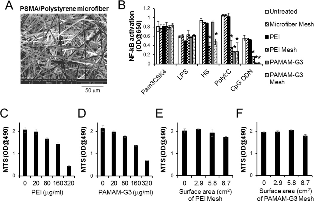
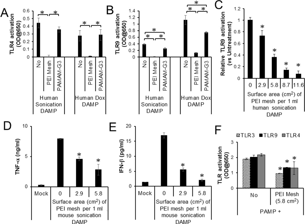
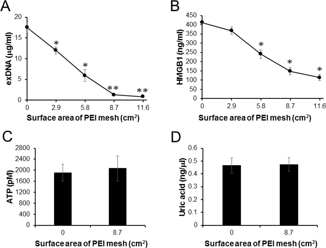
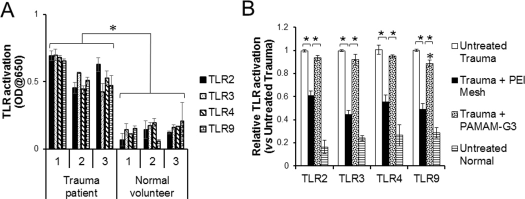
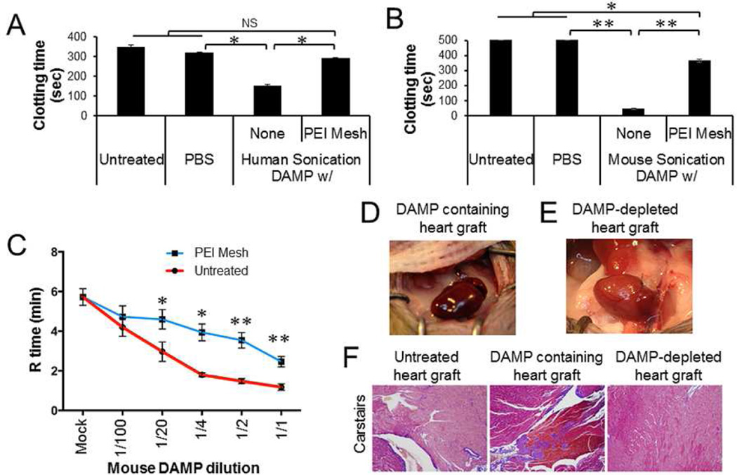
Trauma patients produce a host of danger signals and high levels of damage-associated molecular patterns (DAMPs) after cellular injury and tissue damage. These DAMPs are directly and indirectly involved in the pathogenesis of various inflammatory and thrombotic complications in patients with severe injuries. No effective therapeutic agents for the removal of DAMPs from blood or tissue fluid have been developed. Herein, we demonstrated that nucleic acid binding polymers, e.g., polyethylenimine (PEI) and polyamidoamine dendrimers, immobilized onto electrospun microfiber mesh can effectively capture various DAMPs, such as extracellular DNAs and high mobility group box 1 (HMGB1). Furthermore, treatment with PEI-immobilized microfiber mesh abrogated the ability of DAMPs, released from dead and dying cells in culture or found in patients following traumatic injury, to activate innate immune responses and coagulation in vitro and in vivo. Nucleic acid scavenging microfiber meshes represent an effective strategy to combat inflammation and thrombosis in trauma.

Keywords: Inflammation; Microfiber mesh; Nucleic acid scavenger; Thrombosis; Toll like receptor.


Conflict of interest statement

We have no conflict of interest to declare.

Figures

Fig. 1
Fig. 1
NABP-immobilized PSMA/polystyrene microfiber mesh scavenges multiple TLR agonists without cytotoxicity. A. scanning electron microscope (SEM) image of surface of PSMA/polystyrene microfiber mesh. B. Complete culture media (1 ml) supplemented with TLR agonists Pam3CSK4, LPS, Heparan sulfate (HS), polyI:C or CpG ODN, were incubated for 1 min with or without either PEI- or PAMAM-G3-immobilized microfiber mesh (2.9 cm2) at room temperature. The treatment was repeated once. TLR reporter cells were incubated for 3 days in either untreated or treated complete culture media. For the treatment with free NABPs, TLR reporter cells in the complete culture media containing fetal bovine serum and TLR agonist were directly treated with either PEI (20 µg/ml) or PAMAM-G3 (25 µg/ml). The activation of NF-κB in TLR signaling pathway was determined by a colorimetric enzyme assay. C-F. Human primary fibroblasts were cultured in either complete culture media containing free (C) PEI or free (D) PAMAM-G3 at various concentrations or complete culture media pre-treated with (E) PEI-immobilized mesh or (F) PAMAM-G3-immobilized mesh at various surface areas (0.288 mg/cm2 PEI on mesh; 0.128 mg/cm2 PAMAM-G3 on mesh). After 3 days incubation, cell proliferation was determined by a MTS (3-(4,5-dimethylthiazol-2-yl)-5-(3-carboxymethoxyphenyl)-2-(4-sulfophenyl)-2H–tetrazolium) assay. Error bars are S.D.. * Significant different (P < 0.05), vs untreated group.
Fig. 2
Fig. 2
Inhibition of DAMP and PAMP-mediated TLR activation by PEI-immobilized PSMA/polystyrene microfiber mesh. A, B. DAMPs were generated from human and mouse cells that were killed by either sonication or doxorubicin. (A) TLR4 and (B) TLR9 reporter cell lines were stimulated overnight with human DAMPs (20 % v/v) with or without treatment with either PEI-immobilized mesh (5.8 cm2) or PAMAM-G3 (25 µg/ml). C. TLR9 reporter cells were stimulated with DAMPs pre-treated with PEI-immobilized mesh at various surface sizes. The activation of NF-kB in TLR signaling pathway was determined by a colorimetric enzyme assay. D, E. Mouse macrophage cell line was incubated overnight with mouse DAMPs pre-treated with PEI-immobilized DAMPs at various surface sizes. (D) TNF-α and (E) IFN-β production by the cells were determined by ELISA. F. Bacterial PAMPs were pre-treated with or without PEI-immobilized mesh (5.8 cm2). TLR3, TLR4 and TLR9 reporter cell lines were stimulated overnight with the untreated or treated PAMPs (5% v/v). Error bars are S.D.. * Significant different (P < 0.05), between indicated groups or compared with untreated.
Fig. 3
Fig. 3
PEI-immobilized PSMA/polystyrene microfiber mesh capture and remove exDNA and HMGB1. Human DAMPs generated by sonication-induced cell death were treated with PEI-immobilized PSMA/polystyrene meshes at various surface sizes. The levels of (A) exDNAs, (B) HMGB1, (C) ATP and (D) uric acid in the DAMPs were determined by PicoGreen assay, ELISA, Bioluminescence assay and Fluorometric assay, respectively. Error bars are S.D.. * Significant different (P < 0.05), vs Untreated. ** Significant different (P < 0.01), vs Untreated.
Fig. 4
Fig. 4
PEI-immobilized microfiber mesh inhibits pro-inflammatory DAMPs from trauma patients. A. TLR2, TLR3, TLR4 and TLR9 reporter cells were incubated overnight with sera (20% v/v) isolated from either trauma patients or normal healthy volunteers. B. Undiluted serum was treated with or without either PEI-immobilized microfiber mesh (8.7 cm2) or PAMAM-G3 (25 µg/ml). NF-κB activation in the TLR reporter cell lines was determined by a colorimetric assay. Error bars are S.D.. * P < 0.05. * P < 0.01.
Fig. 5
Fig. 5
Inhibition of DAMP-induced clotting by PEI-immobilized PSMA/polystyrene microfiber mesh. Human and mouse sonication-induced DAMPs were incubated for 1 min with or without PEI-immobilized PSMA/polystyrene microfiber meshes (2.9 cm2). The treatment was repeated four times. A, B. Platelet-depleted plasma from human and mouse normal blood were incubated with the DAMPs (10% v/v) and the clotting time measured. C. Mouse whole blood was incubated with the DAMPs at various dilutions with PBS. The coagulation (R) time was detected by Thromboelastography (TEG). D, E. Donor hearts isolated from normal mice (n=3) was perfused with the DAMP (2 ml), followed by heterotrophic heart transplantation. Heart beating and thrombosis of donor heart was monitored. The image was captured 30 min after unclamping. F. Representative sections from untreated, DAMP-treated, and PEI-immobilized mesh-filtered DAMP-treated allografts harvested 30 min after unclamping. All were stained with Carstairs (modified Masson’s trichrome) to visualize platelets (purple), erythrocytes (clear yellow/orange), and fibrin (Bright red/orange) taken at x100 magnification. Data represent three independent experiments. Error bars are S.D.. NS: statistically non-significant. * P < 0.05. ** P < 0.01.

Comment in

References

    1. Venereau E, Ceriotti C, Bianchi ME. DAMPs from Cell Death to New Life. Frontiers in immunology. 2015;6:422. - PMC - PubMed
    1. Kawai T, Akira S. TLR signaling. Seminars in immunology. 2007;19:24–32. - PubMed
    1. Liaw PC, Ito T, Iba T, Thachil J, Zeerleder S. DAMP and DIC: The role of extracellular DNA and DNA-binding proteins in the pathogenesis of DIC. Blood reviews. 2015 - PubMed
    1. Stoecklein VM, Osuka A, Lederer JA. Trauma equals danger--damage control by the immune system. J Leukoc Biol. 2012;92:539–551. - PMC - PubMed
    1. Leulier F, Lemaitre B. Toll-like receptors--taking an evolutionary approach. Nat Rev Genet. 2008;9:165–178. - PubMed

Publication types