. 2017 Jan 3;8(1):e02120-16.
doi: 10.1128/mBio.02120-16.

Proximity Interactions among Basal Body Components in Trypanosoma brucei Identify Novel Regulators of Basal Body Biogenesis and Inheritance

Affiliations

Proximity Interactions among Basal Body Components in Trypanosoma brucei Identify Novel Regulators of Basal Body Biogenesis and Inheritance

Hung Quang Dang et al. mBio. .

Abstract

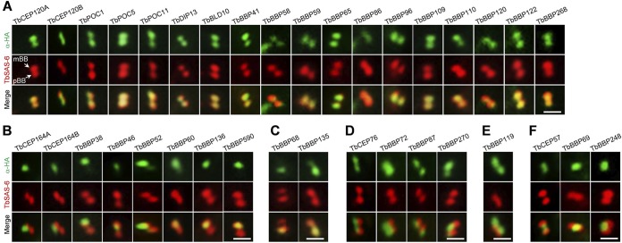
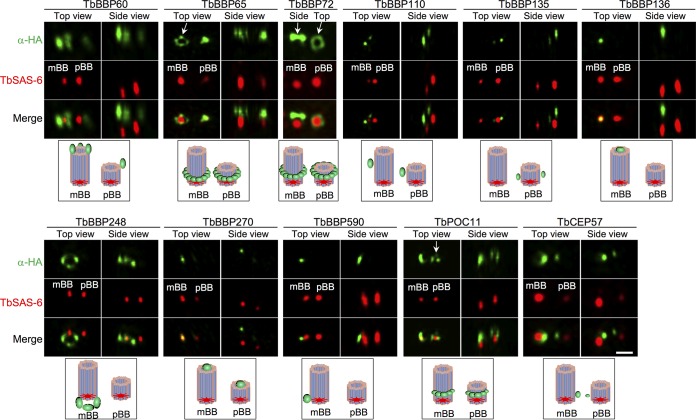
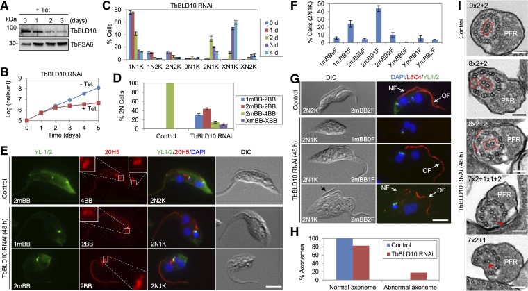
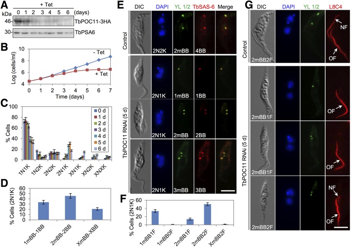
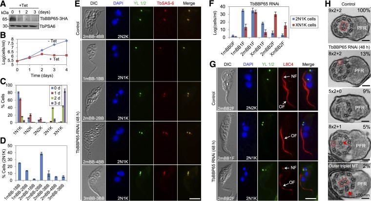
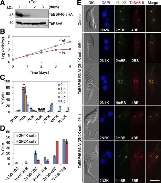
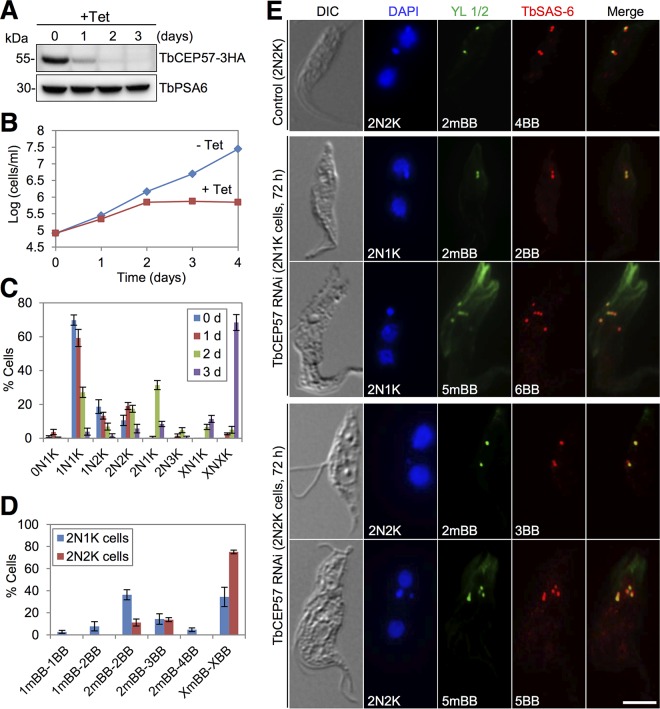
The basal body shares similar architecture with centrioles in animals and is involved in nucleating flagellar axonemal microtubules in flagellated eukaryotes. The early-branching Trypanosoma brucei possesses a motile flagellum nucleated from the basal body that consists of a mature basal body and an adjacent pro-basal body. Little is known about the basal body proteome and its roles in basal body biogenesis and flagellar axoneme assembly in T. brucei Here, we report the identification of 14 conserved centriole/basal body protein homologs and 25 trypanosome-specific basal body proteins. These proteins localize to distinct subdomains of the basal body, and several of them form a ring-like structure surrounding the basal body barrel. Functional characterization of representative basal body proteins revealed distinct roles in basal body duplication/separation and flagellar axoneme assembly. Overall, this work identified novel proteins required for basal body duplication and separation and uncovered new functions of conserved basal body proteins in basal body duplication and separation, highlighting an unusual mechanism of basal body biogenesis and inheritance in this early divergent eukaryote.

Importance: The basal body in the early-branching protozoan Trypanosoma brucei nucleates flagellum assembly and also regulates organelle segregation, cell morphogenesis, and cell division. However, the molecular composition and the assembly process of the basal body remain poorly understood. Here, we identify 14 conserved basal body proteins and 25 trypanosome-specific basal body proteins via bioinformatics, localization-based screening, and proximity-dependent biotin identification. We further localized these proteins to distinct subdomains of the basal body by using fluorescence microscopy and superresolution microscopy, discovered novel regulators of basal body duplication and separation, and uncovered new functions of conserved basal body proteins in basal body duplication and separation. This work lays the foundation for dissecting the mechanisms underlying basal body biogenesis and inheritance in T. brucei.


Figures

FIG 1
FIG 1
Subcellular localizations of basal body proteins. Proteins were endogenously tagged with a triple-HA epitope and detected by coimmunostaining with FITC-conjugated anti-HA MAb and anti-TbSAS-6 polyclonal antibody. (A) Proteins localizing to the vicinity of mBB and pBB. (B) Proteins localizing to the vicinity of mBB. (C) Proteins localizing to the vicinity of pBB. (D) Proteins localizing to the distal ends (flagellar sides) of mBB and pBB. (E) A protein that localizes to the proximal ends (kinetoplast side) of mBB and pBB. (F) Proteins localizing between mBB and pBB. Bars, 5 µm.
FIG 2
FIG 2
3D-SIM superresolution microscopic analysis of basal body protein localization. Cells were coimmunostained with anti-HA MAb and anti-TbSAS-6 polyclonal antibody and visualized using the DeltaVision OMX v 4 Blaze microscope. The cartoon below the microscopy images illustrates the localization of the protein (in green) in the basal body. About 40 cells were imaged for each protein. Note that the different sizes of mBBs and pBBs in some images could be due to the differences in their distances from the camera. Bars, 0.5 µm.
FIG 3
FIG 3
TbBLD10 is required for pro-basal body biogenesis and axoneme assembly. (A) Western blotting with anti-TbBLD10 antibody to monitor the TbBLD10 protein level. Levels of TbPSA6, the T. brucei proteasome subunit alpha-6, served as the loading control. (B) RNAi of TbBLD10 inhibited cell proliferation. (C) Quantification of the numbers of nuclei (N) and kinetoplasts (K) before and after TbBLD10 RNAi. A total of 200 cells were counted for each time point, and error bars indicate standard deviations calculated from three independent experiments. (D) Quantification of cells with different numbers of mBBs and total basal body (mBBs and pBBs) in control 2N2K cells and TbBLD10-deficient 2N1K cells. A total of 200 cells were counted for each cell type, and error bars indicate standard deviations calculated from three independent experiments. (E) Coimmunostaining of cells with YL 1/2 to label mBBs and with 20H5 to label mBBs and pBBs. Bar, 5 µm. (F) Quantification of cells with different numbers of flagella and mBBs in TbBLD10-deficient 2N1K cells. A total of 200 cells were counted, and error bars indicate standard deviations calculated from three independent experiments. (G) Coimmunostaining of cells with L8C4 to label the flagella and YL 1/2 to label mBBs. NF, new flagellum; OF, old flagellum. Bar, 5 µm. (H) Quantification of axonemes with normal or abnormal structures in control and TbBLD10 RNAi cells. A total of 95 sections from control cells and 126 sections from TbBLD10 RNAi cells were counted. (I) Morphology of the axoneme in control and TbBLD10 RNAi cells. Note that the orientation of the central pair (outlined in a red rectangle) in TbBLD10 RNAi cells was altered. The red oval outlines the missing outer doublet in the axoneme of a TbBLD10 RNAi cell. The red brackets show the enlarged distance between the outer doublets in a TbBLD10 RNAi cell. The red open arrowhead indicates an outer singlet in a TbBLD10 RNAi cell. The red arrow indicates a central singlet, instead of a central pair, in a TbBLD10 RNAi cell. Bars, 100 nm.
FIG 4
FIG 4
RNAi of TbPOC11 disrupts pro-basal body biogenesis and flagellum assembly. (A) Western blotting to monitor the protein level of TbPOC11. TbPOC11 was endogenously tagged with a triple-HA epitope in the TbPOC11 RNAi cell line. TbPSA6 served as the loading control. (B) TbPOC11 depletion inhibited cell proliferation. (C) Quantification of cells with different numbers of nuclei (N) and kinetoplasts (K) before and after TbPOC11 RNAi. A total of 200 cells were counted for each time point, and error bars indicate standard deviations calculated from three independent experiments. (D) Quantification of TbPOC11-deficient 2N1K cells with different numbers of mBBs and BBs (both mBBs and pBBs). A total of 200 cells were counted, and error bars indicate standard deviations from three independent experiments. (E) Coimmunostaining of cells with YL 1/2 to label mBBs and anti-TbSAS-6 antibody to label mBBs and pBBs. Bar, 5 µm. (F) Quantification of the numbers of mBBs and flagella in the 2N1K cells from TbPOC11 RNAi. A total of 200 cells were counted, and error bars indicate standard deviations calculated from three independent experiments. (G) Coimmunostaining of cells with YL 1/2 to label mBBs and L8C4 to label the flagella. NF, new flagellum; OF, old flagellum. Bar, 5 µm.
FIG 5
FIG 5
TbBBP65 is required for pro-basal body biogenesis and axoneme assembly. (A) Western blotting to examine the level of TbBBP65. TbBBP65 was endogenously tagged with a triple-HA epitope in the TbBBP65 RNAi cell line. TbPSA6 served as the loading control. (B) TbBBP65 RNAi inhibited cell proliferation. (C) Quantification of cells with different numbers of kinetoplasts (K) and nuclei (N) in control and TbBBP65 RNAi cells. A total of 200 cells were counted for each time point, and error bars indicate standard deviations calculated from three independent experiments. (D) Quantification of TbBBP65-deficient 2N1K cells with different numbers of mBBs and BBs (both mBBs and pBBs). A total of 200 cells were counted, and error bars indicate standard deviations calculated from three independent experiments. (E) Coimmunostaining of cells with YL 1/2 to label mBBs and with anti-TbSAS-6 antibody to label mBBs and pBBs. Bar, 5 µm. (F) Quantification of TbBBP65-deficient 2N1K cells and XN1K (X > 2) cells with different numbers of mBBs and flagella. A total of 200 cells were counted for each cell type, and error bars indicate standard deviations calculated from three independent experiments. (G) Immunostaining of cells with YL 1/2 to label mBBs and with L8C4 to label the flagella. Bar, 5 µm. (H) Morphology of flagellar axonemes in control and TbBBP65 RNAi cells. The red oval outlines a missing outer microtubule doublet in TbBBP65 RNAi cells. The red open arrowhead indicates a central microtubule singlet in a TbBBP65 RNAi cell. The red arrow indicates an outer microtubule triplet in a TbBBP65 RNAi cell. The red rectangle outlines the central microtubule pair in a control cell and a TbBBP65 RNAi cell. Note the orientation of the central pair in the RNAi cell was altered. Bar, 100 nm.
FIG 6
FIG 6
TbBBP46 is required for basal body separation. (A) Western blotting to examine the level of TbBBP46 before and after RNAi induction. TbBBP46 was endogenously tagged with a triple HA epitope in cells harboring the TbBBP46 RNAi construct. TbBBP46-3HA was detected by anti-HA antibody. TbPSA6 served as the loading control. (B) RNAi of TbBBP46 inhibited cell proliferation. (C) Quantification of cells with different numbers of kinetoplasts (K) and nuclei (N) before and after TbBBP46 RNAi. A total of 200 cells were counted for each time point, and error bars indicate standard deviations calculated from three independent experiments. (D) Effect of TbBBP65 RNAi on basal body duplication/separation. Shown is the quantification of the TbBBP46-deficient 2N1K and 2N2K cells with different numbers of mature basal bodies (mBBs) and basal bodies (both mBBs and pBBs). A total of 200 cells were counted, and error bars indicate standard deviations from three independent experiments. (E) Coimmunostaining of control and TbBBP46 RNAi cells with YL 1/2 antibody to label the mature basal body (mBBs) and with anti-TbSAS-6 antibody to label both the mature basal body and pro-basal body. Bar, 5 µm.
FIG 7
FIG 7
TbCEP57 is required for basal body separation and flagellum assembly. (A) Western blotting to monitor the protein level of TbCEP57 before and after TbCEP57 RNAi. TbCEP57 was endogenously tagged with a triple-HA epitope in cells harboring the TbCEP57 RNAi construct. TbPSA6 served as the loading control. (B) TbCEP57 is essential for cell proliferation. (C) Quantification of cells with different numbers of kinetoplasts (K) and nuclei (N) before and after TbCEP57 RNAi. A total of 200 cells were counted for each time point, and error bars indicate standard deviations calculated from three independent experiments. (D) Quantification of cells with different numbers of mature basal bodies (mBBs) and basal bodies (both mBBs and pBBs) in TbCEP57-depleted 2N1K and 2N2K cells. A total of 200 cells were counted for each cell type, and error bars indicate standard deviations calculated from three independent experiments. (E) Immunostaining of control and TbCEP57 RNAi cells with YL 1/2 antibody to label the mature basal body and with anti-TbSAS-6 antibody to label both the mature basal body and pro-basal body. Bar, 5 µm.

Similar articles

Cited by

References

    1. Nakazawa Y, Hiraki M, Kamiya R, Hirono M. 2007. SAS-6 is a cartwheel protein that establishes the 9-fold symmetry of the centriole. Curr Biol 17:2169–2174. doi:10.1016/j.cub.2007.11.046. - DOI - PubMed
    1. Hiraki M, Nakazawa Y, Kamiya R, Hirono M. 2007. Bld10p constitutes the cartwheel-spoke tip and stabilizes the 9-fold symmetry of the centriole. Curr Biol 17:1778–1783. doi:10.1016/j.cub.2007.09.021. - DOI - PubMed
    1. Hodges ME, Scheumann N, Wickstead B, Langdale JA, Gull K. 2010. Reconstructing the evolutionary history of the centriole from protein components. J Cell Sci 123:1407–1413. doi:10.1242/jcs.064873. - DOI - PMC - PubMed
    1. Carvalho-Santos Z, Machado P, Branco P, Tavares-Cadete F, Rodrigues-Martins A, Pereira-Leal JB, Bettencourt-Dias M. 2010. Stepwise evolution of the centriole-assembly pathway. J Cell Sci 123:1414–1426. doi:10.1242/jcs.064931. - DOI - PubMed
    1. Schmidt TI, Kleylein-Sohn J, Westendorf J, Le Clech M, Lavoie SB, Stierhof YD, Nigg EA. 2009. Control of centriole length by CPAP and CP110. Curr Biol 19:1005–1011. doi:10.1016/j.cub.2009.05.016. - DOI - PubMed

Publication types

LinkOut - more resources