. 2017 Jan 3;15(1):1.
doi: 10.1186/s12951-016-0241-6.

Intracellular trafficking and cellular uptake mechanism of PHBV nanoparticles for targeted delivery in epithelial cell lines

Affiliations

Intracellular trafficking and cellular uptake mechanism of PHBV nanoparticles for targeted delivery in epithelial cell lines

Juan P Peñaloza et al. J Nanobiotechnology. .

Abstract

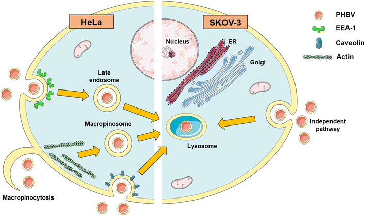
Background: Nanotechnology is a science that involves imaging, measurement, modeling and a manipulation of matter at the nanometric scale. One application of this technology is drug delivery systems based on nanoparticles obtained from natural or synthetic sources. An example of these systems is synthetized from poly(3-hydroxybutyrate-co-3-hydroxyvalerate), which is a biodegradable, biocompatible and a low production cost polymer. The aim of this work was to investigate the uptake mechanism of PHBV nanoparticles in two different epithelial cell lines (HeLa and SKOV-3).

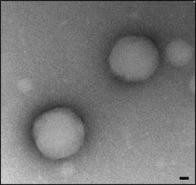
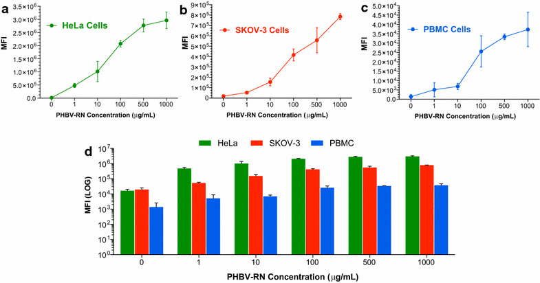
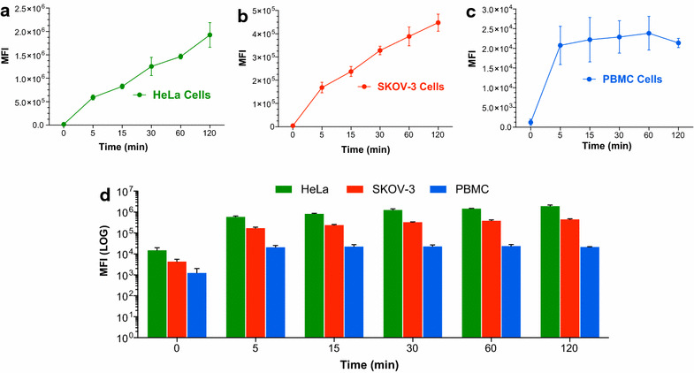
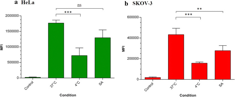
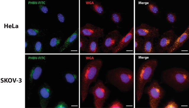
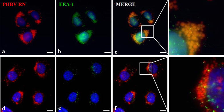
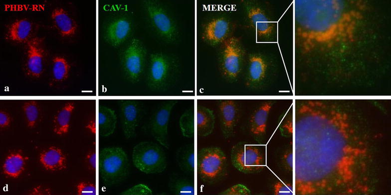
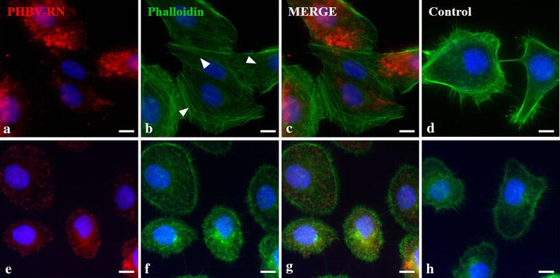
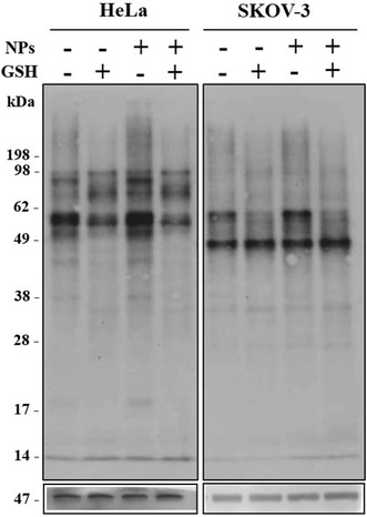
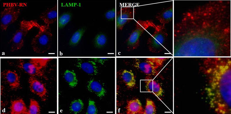
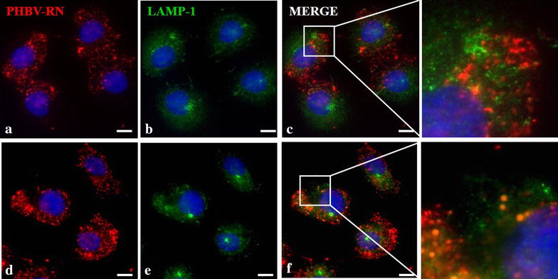
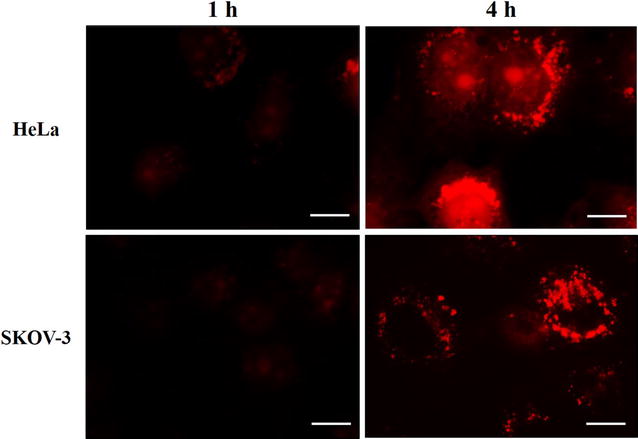
Results: As a first step, we characterized size, shape and surface charge of nanoparticles using dynamic light scattering and transmission electron microscopy. Intracellular incorporation was evaluated through flow cytometry and fluorescence microscopy using intracellular markers. We concluded that cellular uptake mechanism is carried out in a time, concentration and energy dependent way. Our results showed that nanoparticle uptake displays a cell-specific pattern, since we have observed different colocalization in two different cell lines. In HeLa (Cervical cancer cells) this process may occur via classical endocytosis pathway and some internalization via caveolin-dependent was also observed, whereas in SKOV-3 (Ovarian cancer cells) these patterns were not observed. Rearrangement of actin filaments showed differential nanoparticle internalization patterns for HeLa and SKOV-3. Additionally, final fate of nanoparticles was also determined, showing that in both cell lines, nanoparticles ended up in lysosomes but at different times, where they are finally degraded, thereby releasing their contents.

Conclusions: Our results, provide novel insight about PHBV nanoparticles internalization suggesting that for develop a proper drug delivery system is critical understand the uptake mechanism.


Conflict of interest statement

The authors declare that they have no competing interests.

Figures

Fig. 1
Fig. 1
PHBV nanoparticle micrograph. Representative image of PHBV nanoparticles (naked) by transmission electron microscopy (TEM). Objective ×49.000 magnification. Bar size 35 nm
Fig. 2
Fig. 2
Quantification of the cellular uptake of PHBV nanoparticles: time dependency. To evaluate time dependency in the cellular uptake of NPs, HeLa (a), SKOV-3 (b) and PBMC (c) cells were incubated with a 100 µg/mL PHBV-RN at several times. d Mean fluorescence intensity analysis of the three different cells. Mean fluorescence intensity was determined by flow cytometry (n = 3)
Fig. 3
Fig. 3
Quantification of the cellular uptake of PHBV nanoparticles. Concentration dependency. To evaluate concentration dependency in the cellular uptake of NPs, HeLa (a), SKOV-3 (b) and PBMC (c) were incubated with a PHBV-RN solution at different concentrations (1, 10, 100, 500 and 1000 µg/mL) for 2 h. d Mean fluorescence intensity analysis of the three different cells. Mean fluorescence intensity was determined through flow cytometry (n = 3)
Fig. 4
Fig. 4
Quantification of the cellular uptake of PHBV nanoparticles: energy dependency. To evaluate energy dependency in the cellular uptake of NPs, HeLa (a) and SKOV-3 cells (b) were incubated with 100 µg/mL PHBV-RN under different conditions (37, 4 °C, sodium azide). Mean fluorescence intensity was determined through flow cytometry (n = 3). One-way ANOVA and Bonferroni statistical test were used (**p < 0.01, ***p < 0.001, ****p < 0.0001)
Fig. 5
Fig. 5
Characterization of the endocytosis mechanism using WGA marker. HeLa and SKOV-3 cells were incubated with PHBV-FITC for 5 min at 37 °C, in the presence of Alexa Fluor® 555-conjugated WGA. Later, cells were fixed and observed by fluorescence microscopy. Hoechst 33342 was used as a nuclear stain. Objective: ×60 magnification. Bar size 10 µm
Fig. 6
Fig. 6
Characterization of the endocytosis mechanism using EEA-1 marker. HeLa (ac) and SKOV-3 (df) cells were incubated with PHBV-RN nanoparticles for 15 min at 37 °C; then cells were fixed and permeabilized. Later, an immunofluorescence was performed using anti-EEA1 (1:100) and anti-mouse Alexa Fluor® 488 (1:500) as primary and secondary antibody respectively. Finally, cells were observed through fluorescence microscopy. Hoechst 33342 was used as nuclear stain. Objective: ×60 magnification. Bar size 10 µm
Fig. 7
Fig. 7
Characterization of the endocytosis mechanism using CAV-1 marker. HeLa (ac) and SKOV-3 (df) cells were incubated with PHBV-RN nanoparticles for 15 min at 37 °C, then cells were fixed and permeabilized. Later, an immunofluorescence was performed using anti-CAV1 (1:100) and anti-mouse Alexa Fluor® 488 (1:500) as primary and secondary antibody respectively. Finally, cells were observed through fluorescence microscopy. Hoechst 33342 was used as nuclear stain. Objective: ×60 magnification. Bar size 10 µm
Fig. 8
Fig. 8
Evaluation of actin filaments rearrangement in the presence of PHBV nanoparticles. HeLa (ad) and SKOV-3 (eh) cells were incubated with PHBV-RN for 15 and 30 min at 37 °C; then cells were permeabilized and incubated with Phalloidin Alexa Fluor® 488 conjugated. Hoechst 33242 was used as nuclear stain. Objective: ×60 magnification. Bar size 10 µm
Fig. 9
Fig. 9
Analysis of proteins involved in cellular uptake of PHBV nanoparticles. HeLa and SKOV-3 cells were subjected to reducible biotin assay to determine possible membrane proteins involved in nanoparticles uptake. 40 µg of proteins from cell lysate were resolved through SDS-PAGE, then transferred to PDVF membrane and finally, incubated with streptavidin-HRP (1:1000). β-actin (47 kDa) was used as loading control
Fig. 10
Fig. 10
Determination of final nanoparticle fate in HeLa cells. HeLa cells were incubated with PHBV-RN nanoparticles for 15 min (ac) and 1 h (df) at 37 °C; then cells were fixed and permeabilized. Later, an immunofluorescence was performed using anti-LAMP1 (1:100) and anti-mouse Alexa Fluor® 488 (1:500) as primary and secondary antibody respectively. Finally, cells were observed through fluorescence microscopy. Hoechst 33342 was used as nuclear stain. Objective: ×60 magnification. Bar size 10 µm
Fig. 11
Fig. 11
Determination of final nanoparticle fate in SKOV-3 cells. SKOV-3 cells were incubated with PHBV-RN nanoparticles for 15 min (ac) and 1 h (df) at 37 °C, then cells were fixed and permeabilized. Later, an immunofluorescence was performed using anti-LAMP1 (1:100) and anti-mouse Alexa Fluor® 488 (1:500) as primary and secondary antibody respectively. Finally, cells were observed through fluorescence microscopy. Hoechst 33342 was used as nuclear stain. Objective: ×60 magnification. Bar size 10 µm
Fig. 12
Fig. 12
Evaluation of nanoparticle degradation. HeLa and SKOV-3 cells were incubated with PHBV-Lysotracker® DND-99/nanoparticles for 4 h at 37 °C, then cells were fixed and observed through fluorescence microscopy. Objective: ×60 magnification. Bar size 10 µm
Fig. 13
Fig. 13
PHBV nanoparticle intracellular trafficking pathway in HeLa and SKOV-3 cells scheme. Our results showed different endocytosis pathway depending on the cell context but the same final destination

References

    1. Wilczewska AZ, Niemirowicz K, Markiewicz KH, Car H. Nanoparticles as drug delivery systems. Pharmacol Rep. 2012;64:1020–1037. doi: 10.1016/S1734-1140(12)70901-5. - DOI - PubMed
    1. De Jong WH. Drug delivery and nanoparticles : applications and hazards. Int J Nanomed. 2008;3:133–149. doi: 10.2147/IJN.S596. - DOI - PMC - PubMed
    1. Chan JM, Valencia PM, Zhang L, Langer R, Farokhzad OC. Polymeric nanoparticles for drug delivery. 2010:163–175. doi:10.1007/978-1-60761-609-2_11. - PubMed
    1. Grabrucker AM, Garner CC, Boeckers TM, Bondioli L, Ruozi B, Forni F, Vandelli MA, Tosi G. Development of novel Zn 2+ loaded nanoparticles designed for cell-type targeted drug release in CNS neurons: in vitro evidences. PLoS ONE. 2011 - PMC - PubMed
    1. Wang M, Hu H, Sun Y, Qiu L, Zhang J, Guan G, Zhao X, Qiao M, Cheng L, Cheng L, Chen D. Biomaterials A pH-sensitive gene delivery system based on folic acid-PEG-chitosan e PAMAM-plasmid DNA complexes for cancer cell targeting. Biomaterials. 2013;34:10120–10132. doi: 10.1016/j.biomaterials.2013.09.006. - DOI - PubMed

Substances