

The abnormal expression of oxytocin receptors in the uterine junctional zone in women with endometriosis
- PMID: 28049501
- PMCID: PMC5209923
- DOI: 10.1186/s12958-016-0220-7
The abnormal expression of oxytocin receptors in the uterine junctional zone in women with endometriosis
Abstract
Background: The junctional zone (JZ), also called as the endometrial-myometrial junction, is related to peristaltic-like movements in the non-pregnant uterus. Hyperperistalsis and dysperistalsis of uterus constructions might underlie many important disorders such as dysmenorrhea, infertility, endometriosis, implantation failure. The major proteins for uterine contraction of the non-pregnant uterus may be Oxytocin (OT) and oxytocin receptor (OTR). The objective of this study was to inspect the expression of OTR in isthmic and mid-fundal parts of the uterine junctional zone at different stages of the follicular cycle in patients with and without endometriosis.
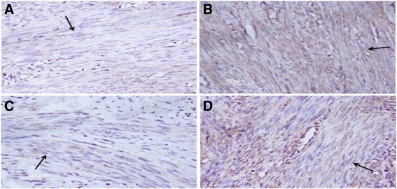
Methods: Uterine biopsies containing endometrium and junctional zone were collected from the isthmic and mid-fundal parts of the anterior wall after hysterectomy. The OTR expression was evaluated by immunohistochemistry.
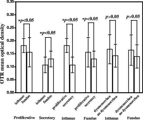
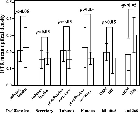
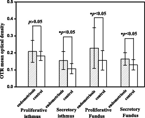
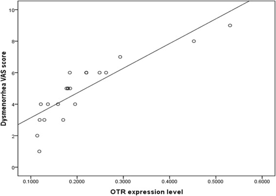
Results: In the control uterus, OTR expression in the isthmic region was significantly higher than in the fundal region in the proliferative phase (p < 0.05) but significantly lower in the secretory phase (p < 0.05). And the expression of OTR in the proliferative phase was significantly higher than that in the secretory phase in both isthmic and fundal regions (p = 0.000 and 0.049, respectively). However, in endometriosis uteri, OTR expression in the isthmic region showed no significant difference with that in the fundal region in both proliferative and secretory phases (p = 0.597 and 0.736, respectively). In both isthmic and fundal regions, OTR expression was not significantly different between the proliferative phase and secretory phase (p = 0.084 and 0.222, respectively). OTR expression in fundal regions of revised ASRM I and II endometriosis were lower than that of revised ASRM III and IV (p = 0.049). In the fundal region of JZ, the expression of OTR in ovarian endometriosis was significantly lower than that in deep infiltrating endometriosis (p = 0.046). The expression level of OTR in the funds region is positively associated with the severity of dysmenorrhea in endometriosis group (r = 0.870, p < 0.05). Comparing to normal uteri, the expression of OTR in the secretory phase was significantly higher in the endometriosis uteri (p < 0.05). In the fundus of endometriosis uteri, OTR expression was significantly higher in both the proliferative and secretory phases (p = 0.045 and 0.028, respectively).
Conclusion: OTR expression in the JZ of women with endometriosis changes significantly, which may result in abnormal uterine contractile activity, reducing the endometriosis-related fertility and dysmenorrhea.
Keywords: Endometriosis; Junctional zone; Oxytocin receptor.
Figures

Similar articles
-
Expression of oxytocin receptors in the uterine junctional zone in women with adenomyosis.Acta Obstet Gynecol Scand. 2015 Apr;94(4):412-8. doi: 10.1111/aogs.12595. Epub 2015 Mar 1. Acta Obstet Gynecol Scand. 2015. PMID: 25627343
-
[Relationship between ultrastructural features with the expression of connexin 43 in the uterine junction zone and pathogenesis of adenomyosis].Zhonghua Fu Chan Ke Za Zhi. 2010 Oct;45(10):762-6. Zhonghua Fu Chan Ke Za Zhi. 2010. PMID: 21176558 Chinese.
-
Possible roles of oxytocin receptor and vasopressin-1α receptor in the pathomechanism of dysperistalsis and dysmenorrhea in patients with adenomyosis uteri.Fertil Steril. 2010 Dec;94(7):2541-6. doi: 10.1016/j.fertnstert.2010.03.015. Epub 2010 Apr 22. Fertil Steril. 2010. PMID: 20413116
-
Myometrial oxytocin receptor expression and intracellular pathways.Minerva Ginecol. 2014 Jun;66(3):267-80. Minerva Ginecol. 2014. PMID: 24971782 Review.
-
The uterine peristaltic pump. Normal and impeded sperm transport within the female genital tract.Adv Exp Med Biol. 1997;424:267-77. Adv Exp Med Biol. 1997. PMID: 9361805 Review.
Cited by
-
Perioperative Suppression of Schwann Cell Dedifferentiation Reduces the Risk of Adenomyosis Resulting from Endometrial-Myometrial Interface Disruption in Mice.Biomedicines. 2022 May 24;10(6):1218. doi: 10.3390/biomedicines10061218. Biomedicines. 2022. PMID: 35740240 Free PMC article.
-
Archimetrosis: the evolution of a disease and its extant presentation : Pathogenesis and pathophysiology of archimetrosis (uterine adenomyosis and endometriosis).Arch Gynecol Obstet. 2023 Jan;307(1):93-112. doi: 10.1007/s00404-022-06597-y. Epub 2022 May 21. Arch Gynecol Obstet. 2023. PMID: 35596746 Free PMC article.
-
Effect of monosodium glutamate on serum sex hormones and uterine histology in female rats along with its molecular docking and in-silico toxicity.Heliyon. 2022 Oct 5;8(10):e10967. doi: 10.1016/j.heliyon.2022.e10967. eCollection 2022 Oct. Heliyon. 2022. PMID: 36237979 Free PMC article.
-
Analysis of the effect of phloroglucinol on pregnancy outcomes involving frozen embryo transfers in patients with endometriosis: A retrospective case-control study.Front Surg. 2023 Jan 6;9:994775. doi: 10.3389/fsurg.2022.994775. eCollection 2022. Front Surg. 2023. PMID: 36684314 Free PMC article.
-
Plasma Oxytocin Concentrations During and After Gestation in Japanese Pregnant Women Affected by Anxiety Disorder and Endometriosis.Yonago Acta Med. 2020 Nov 5;63(4):301-307. doi: 10.33160/yam.2020.11.012. eCollection 2020 Nov. Yonago Acta Med. 2020. PMID: 33253338 Free PMC article.
References
MeSH terms
Substances
LinkOut - more resources
Full Text Sources
Other Literature Sources
Medical
