Prepancreatic postduodenal portal vein: a case report and review of the literature
- PMID: 28049511
- PMCID: PMC5209868
- DOI: 10.1186/s13256-016-1165-3
Prepancreatic postduodenal portal vein: a case report and review of the literature
Abstract
Background: Prepancreatic postduodenal portal vein is extremely rare, with only 13 cases reported in the literature.
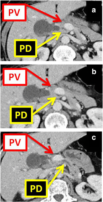
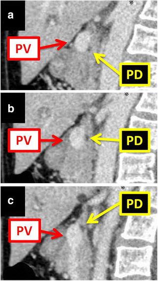
Case presentation: A 55-year-old white woman presented to our emergency department with abdominal pain. She underwent a computed tomography of her abdomen, which showed a portal vein coursing anterolaterally to her pancreas and posteriorly to the first portion of her duodenum, constituting a prepancreatic postduodenal portal vein. Imaging revealed choledocholithiasis, requiring endoscopic sphincterotomy, but due to a history of a gastric bypass procedure, she was lost to follow-up after being referred to an advanced endoscopist. This represents the 14th reported case of prepancreatic postduodenal portal vein.
Conclusions: Awareness of this rare anomaly is paramount, and will help surgeons and interventional radiologists to avoid complications related to inadvertent injury to the portal vein, which could be life-threatening.
Keywords: Anomaly; Case report; Literature review; Pancreatic surgery; Portal vein.
Figures

Similar articles
-
Hepatobiliary-pancreatic surgery for patients with a prepancreatic postduodenal portal vein: a case report and literature review.BMC Surg. 2022 Feb 13;22(1):55. doi: 10.1186/s12893-022-01508-z. BMC Surg. 2022. PMID: 35152891 Free PMC article. Review.
-
Prepancreatic postduodenal portal vein: a new hypothesis for the development of the portal venous system.Jpn J Radiol. 2010 Feb;28(2):157-61. doi: 10.1007/s11604-009-0386-4. Epub 2010 Feb 26. Jpn J Radiol. 2010. PMID: 20182851
-
Prepancreatic postduodenal portal vein: a rare vascular variant detected on imaging.Surg Radiol Anat. 2013 Sep;35(7):631-4. doi: 10.1007/s00276-013-1081-9. Epub 2013 Feb 8. Surg Radiol Anat. 2013. PMID: 23392551 Review.
-
Prepancreatic postduodenal portal vein: report of a case.Surg Today. 2003;33(12):956-9. doi: 10.1007/s00595-003-2601-8. Surg Today. 2003. PMID: 14669093
-
Prepancreatic postduodenal portal vein: a case report and literature review.Surg Case Rep. 2023 Apr 23;9(1):63. doi: 10.1186/s40792-023-01644-5. Surg Case Rep. 2023. PMID: 37087704 Free PMC article.
Cited by
-
Anomalous duplication of the portal vein with prepancreatic postduodenal portal vein.J Rural Med. 2022 Oct;17(4):259-261. doi: 10.2185/jrm.2022-009. Epub 2022 Oct 22. J Rural Med. 2022. PMID: 36397802 Free PMC article.
-
A potential intraoperative disaster averted: Preduodenal portal vein compressing a preduodenal common bile duct.Ann Hepatobiliary Pancreat Surg. 2021 Feb 28;25(1):145-149. doi: 10.14701/ahbps.2021.25.1.145. Ann Hepatobiliary Pancreat Surg. 2021. PMID: 33649268 Free PMC article.
-
Hepatobiliary-pancreatic surgery for patients with a prepancreatic postduodenal portal vein: a case report and literature review.BMC Surg. 2022 Feb 13;22(1):55. doi: 10.1186/s12893-022-01508-z. BMC Surg. 2022. PMID: 35152891 Free PMC article. Review.
-
Pancreaticoduodenectomy performed for a patient with prepancreatic postduodenal portal vein: a case report and literature review.Front Med (Lausanne). 2023 Aug 16;10:1180759. doi: 10.3389/fmed.2023.1180759. eCollection 2023. Front Med (Lausanne). 2023. PMID: 37654663 Free PMC article.
-
Prepancreatic postduodenal portal vein discovered in a pediatric patient undergoing total pancreatectomy with islet autotransplantation: a case report and review of literature.Front Surg. 2025 Jan 7;11:1509807. doi: 10.3389/fsurg.2024.1509807. eCollection 2024. Front Surg. 2025. PMID: 39840268 Free PMC article.
References
-
- Shimizu D, Fujii T, Suenaga M, Niwa Y, Okumura N, Kanda M, et al. A case of carcinoma of the ampulla of Vater with anomaly of the portal venous system: prepancreatic postduodenal portal vein. Jpn J Gastroenterol Surg. 2014;47:275–80. doi: 10.5833/jjgs.2013.0229. - DOI
-
- Jung YJ, Lee SJ, Yang SB, Park WK, Chang JC, Kim JW, et al. Prepancreatic postduodenal portal vein: a case report. J Korean Radiol Soc. 2005;53:435–9. doi: 10.3348/jkrs.2005.53.6.435. - DOI
-
- Tanaka K, Sano K, Yano F, Oohira Y, Takahashi T, Suda K. A case of carcinoma of the inferior bile duct with anomaly of the portal venous system — prepancreatic, postduodenal portal vein. Operation. 2000;54:1147–50.
-
- Ozeki Y, Tateyama K, Sumi Y, Yamada T, Yamauchi K, Bando M. Major hepatectomy for liver tumor with anomalous portal branching. Jpn J Gastroenterol Surg. 1999;32:2301–5. doi: 10.5833/jjgs.32.2301. - DOI
-
- Yasui M, Tsunoo H, Nakahara H, Asano M, Fujita H. Portal vein positioned anterior to the pancreas and posterior to the duodenum – report of a case. J Jpn Surg Assoc. 1998;59:526–31.
Publication types
MeSH terms
LinkOut - more resources
Full Text Sources
Other Literature Sources
Medical

