Review
. 2017 Jan 3;9(1):a027706.
doi: 10.1101/cshperspect.a027706.

Regulating the Suprachiasmatic Nucleus (SCN) Circadian Clockwork: Interplay between Cell-Autonomous and Circuit-Level Mechanisms

Affiliations
Review

Regulating the Suprachiasmatic Nucleus (SCN) Circadian Clockwork: Interplay between Cell-Autonomous and Circuit-Level Mechanisms

Erik D Herzog et al. Cold Spring Harb Perspect Biol. .

Abstract

The suprachiasmatic nucleus (SCN) is the principal circadian clock of the brain, directing daily cycles of behavior and physiology. SCN neurons contain a cell-autonomous transcription-based clockwork but, in turn, circuit-level interactions synchronize the 20,000 or so SCN neurons into a robust and coherent daily timer. Synchronization requires neuropeptide signaling, regulated by a reciprocal interdependence between the molecular clockwork and rhythmic electrical activity, which in turn depends on a daytime Na+ drive and nighttime K+ drag. Recent studies exploiting intersectional genetics have started to identify the pacemaking roles of particular neuronal groups in the SCN. They support the idea that timekeeping involves nonlinear and hierarchical computations that create and incorporate timing information through the interactions between key groups of neurons within the SCN circuit. The field is now poised to elucidate these computations, their underlying cellular mechanisms, and how the SCN clock interacts with subordinate circadian clocks across the brain to determine the timing and efficiency of the sleep-wake cycle, and how perturbations of this coherence contribute to neurological and psychiatric illness.


Figures

Figure 1.
Figure 1.
Isolated neurons of the suprachiasmatic nucleus (SCN) are competent, cell-autonomous circadian pacemakers. (A,B) Micrographs of cultured SCN neurons before A, and after B, physically isolating a single neuron (arrow). Scale bars, 50 μm. (C) Recording of PER2 expression using a bioluminescent reporter of PER2 abundance reveals persistent daily rhythms before and after the neuron was isolated. (From Webb et al. 2009; reproduced, with permission, from the authors.)
Figure 2.
Figure 2.
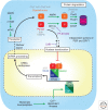
Schematic diagram of the intracellular suprachiasmatic nucleus (SCN) clockwork with points of regulation by mutations and drugs. The canonical clockwork involves a transcription–translational negative feedback loop in which PER–CRY dimers inhibit their own transcription by repressing the actions of CLOCK–BMAL1 dimers on E-box elements in clock genes. Beyond this, mRNA maturation and posttranslational regulation of clock gene products including phosphorylation by AMPK and CK1, ubiquitinylation by FBXL3, FBXL21, and βTrCP, and translational regulation by eIF4E and mRNA methylation contribute to oscillation and determine circadian period. Deletions of Cry1 or Cry2, circadian mutations (gray) in PER genes (Edo and FASP), CK1ɛ (Tau), Fbxl3 (Afterhours, Overtime), and CLOCK (Δ19) as well as drug manipulations (purple, Leptomycin B and PF-670462) highlight key points of regulatory control of the molecular clock.
Figure 3.
Figure 3.
Temporal and spatial circadian programs in the suprachiasmatic nucleus (SCN): calcium and gene expression. (A) Representative images from combined bioluminescent and fluorescent recordings of circadian PER2 (cyan) and [Ca2+]i (green) in organotypic SCN slice culture. (B) Schematic plot of “A day in the life of the SCN” assembled from a series of combined recordings as in A, registered via PER2 and [Ca2+]i rhythms. This reveals phase-specific circadian cycles of electrical activity, calcium-dependent gene expression (CRE), and transcriptional and posttranslational feedback loop (TTFL) functions, clustered around circadian day, and electrical inactivity, and peroxiredoxin overoxidation in circadian night. The electrical activity peak and molecular cycle are intimately interdependent. (C) Circuit-level SCN timekeeping is embodied in a spatiotemporal wave of circadian gene expression, plotted as the center of mass of PER2 bioluminescent signal over three sequential circadian cycles of wild-type (WT) SCN (left) (overlaid with a standardized SCN schematic). This structure is lost in CRY-deficient SCN (right). (Redrawn from data in Edgar et al. 2012, Brancaccio et al. 2013, and Edwards et al. 2016.)
Figure 4.
Figure 4.
Circadian regulation of suprachiasmatic nucleus (SCN) neuronal excitability. A schematic that summarizes the daily changes in excitability of SCN neurons as a “bicycle model.” Each neuron has the intrinsic capacity to generate daily oscillations in electrical activity. During the day or “up-state,” a Na+ leak conductance (gNALCN) increases while the overall K+ conductance (gK) decreases, thereby resulting in an increase in input resistance (RIN) and a more depolarized membrane potential (Vm) and higher firing rates. At night, the neurons enter a “down-state,” with lower RIN, hyperpolarized Vm, elevated gK, and lower gNALCN and firing rates. In this way, the excitatory drive of Na-currents during the day is opposed by elevated K-currents at night. (Redrawn from data in Flourakis et al. 2015.)
Figure 5.
Figure 5.
Vasoactive intestinal peptide (VIP) as a circadian synchronizer and de-synchronizer of suprachiasmatic nucleus (SCN) circuits. (A) Aggregate PER2 bioluminescence from VIP-null SCN recorded before and after graft with wild-type SCN (arrow). In the absence of VIP, SCN organotypic slices lose circadian amplitude and the coherence of cell-autonomous molecular oscillations, and these can be restored by coculture with a wild-type, VIP-competent SCN. (B) Raster plots of cellular PER2 bioluminescence recorded by CCD, before and after grafting (different slice from A). (C) Rayleigh plots of cellular phase determined in relation to grafting. (D) In wild-type SCN, treatment with exogenous VIP can desynchronize the previously tightly coupled circuit. (E, F) Raster and Rayleigh plots as in B and C. (Redrawn from data in Maywood et al. 2011b and An et al. 2013.)
Figure 6.
Figure 6.
Dissection of suprachiasmatic nucleus (SCN) circuit-level pacemaking by intersectional genetic approaches. (A) By manipulating components of the transcriptional and posttranslational feedback loop (TTFL) (BMAL, CLOCK) or the TTFL regulator, CK1ɛ, in defined subpopulations of SCN neurons (AVP, NMS, D1aR), it is possible to alter the period and/or the coherence of mouse activity/rest behavioral rhythms. (Redrawn from data in Lee et al. 2015, Mieda et al. 2015, and Smyllie et al. 2016.) (B) Schematic representation (left) of the partially overlapping subpopulations of SCN neurons, and a meta-analysis (right) of putative pace-setting regions. (Redrawn from data in Smyllie et al. 2016.)

References

    1. Abel JH, Meeker K, Granados-Fuentes D, St John PC, Wang TJ, Bales BB, Doyle FJ III, Herzog ED, Petzold LR. 2016. Functional network inference of the suprachiasmatic nucleus. Proc Natl Acad Sci 113: 4512–4517. - PMC - PubMed
    1. Albus H, Bonnefont X, Chaves I, Yasui A, Doczy J, van der Horst GT, Meijer JH. 2002. Cryptochrome-deficient mice lack circadian electrical activity in the suprachiasmatic nuclei. Curr Biol 12: 1130–1133. - PubMed
    1. Albus H, Vansteensel MJ, Michel S, Block GD, Meijer JH. 2005. A GABAergic mechanism is necessary for coupling dissociable ventral and dorsal regional oscillators within the circadian clock. Curr Biol 15: 886–893. - PubMed
    1. Allen CN, Nitabach MN, Colwell CS. 2016. Membrane currents, gene expression, and circadian clocks. Cold Spring Harb Perspect Biol 10.1101/cshperspect.a027714. - DOI - PMC - PubMed
    1. An S, Irwin RP, Allen CN, Tsai C, Herzog ED. 2011. Vasoactive intestinal polypeptide requires parallel changes in adenylate cyclase and phospholipase C to entrain circadian rhythms to a predictable phase. J Neurophysiol 105: 2289–2296. - PMC - PubMed

LinkOut - more resources