. 2017 Jan 3;10(460):eaag1064.
doi: 10.1126/scisignal.aag1064.

RETRACTED: The receptor tyrosine kinase AXL mediates nuclear translocation of the epidermal growth factor receptor

Affiliations

RETRACTED: The receptor tyrosine kinase AXL mediates nuclear translocation of the epidermal growth factor receptor

Toni M Brand et al. Sci Signal. .

Retraction in

  • Retraction.
    Brand TM, Iida M, Corrigan KL, Braverman CM, Coan JP, Flanigan BG, Stein AP, Salgia R, Rolff J, Kimple RJ, Wheeler DL. Brand TM, et al. Sci Signal. 2021 Nov 9;14(708):eabn0168. doi: 10.1126/scisignal.abn0168. Epub 2021 Nov 9. Sci Signal. 2021. PMID: 34752142 Free PMC article. No abstract available.

Abstract

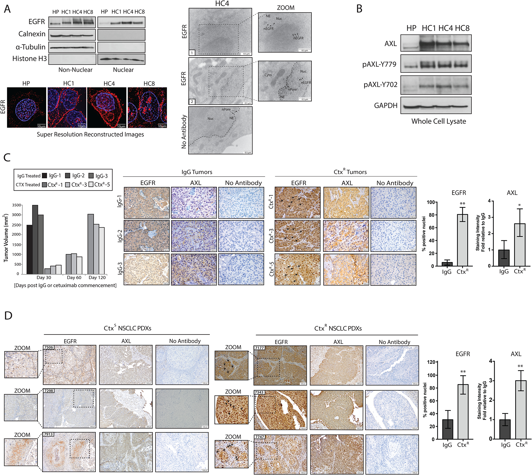
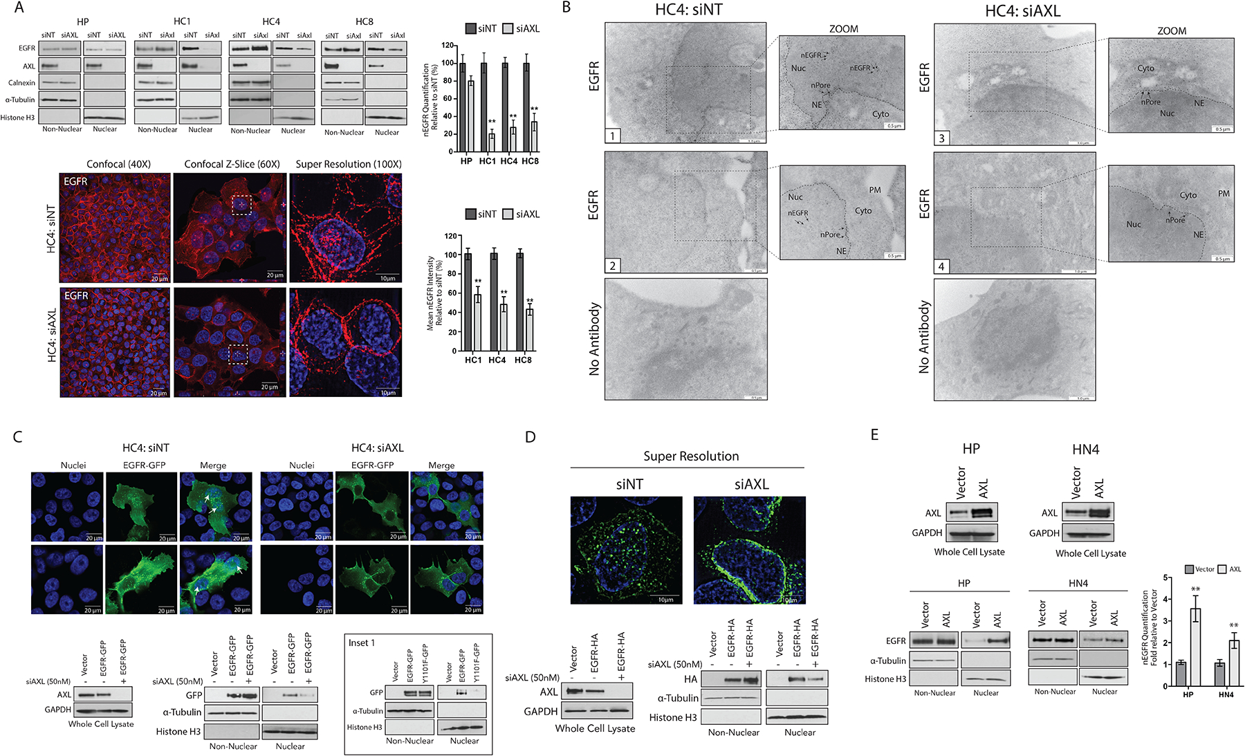
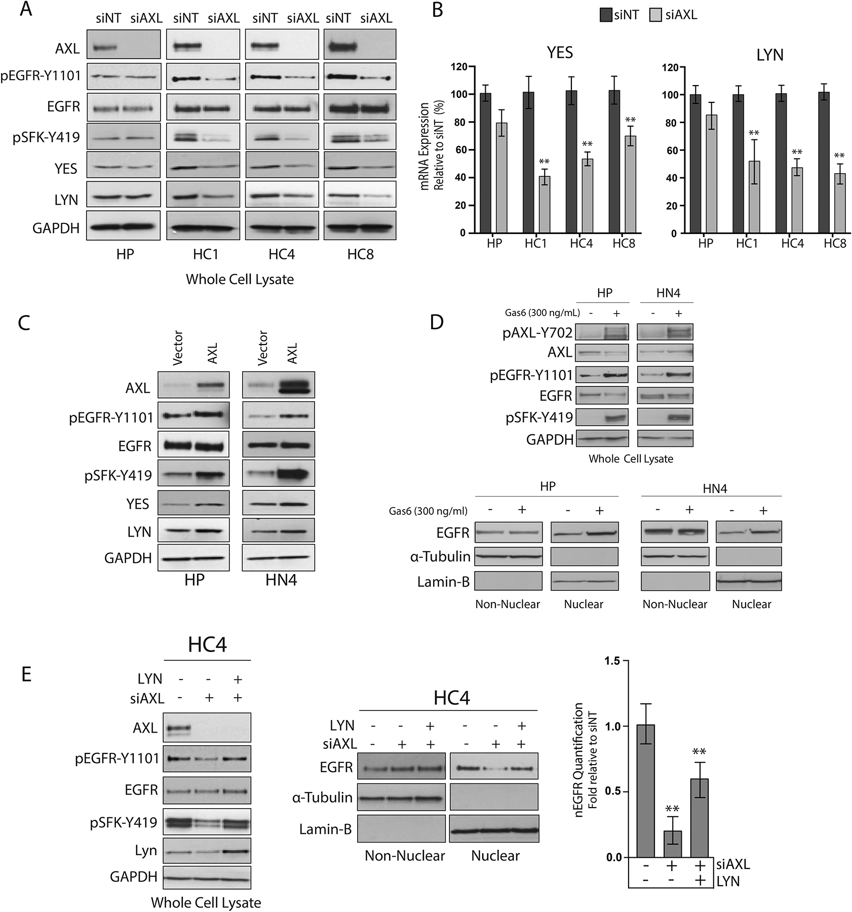
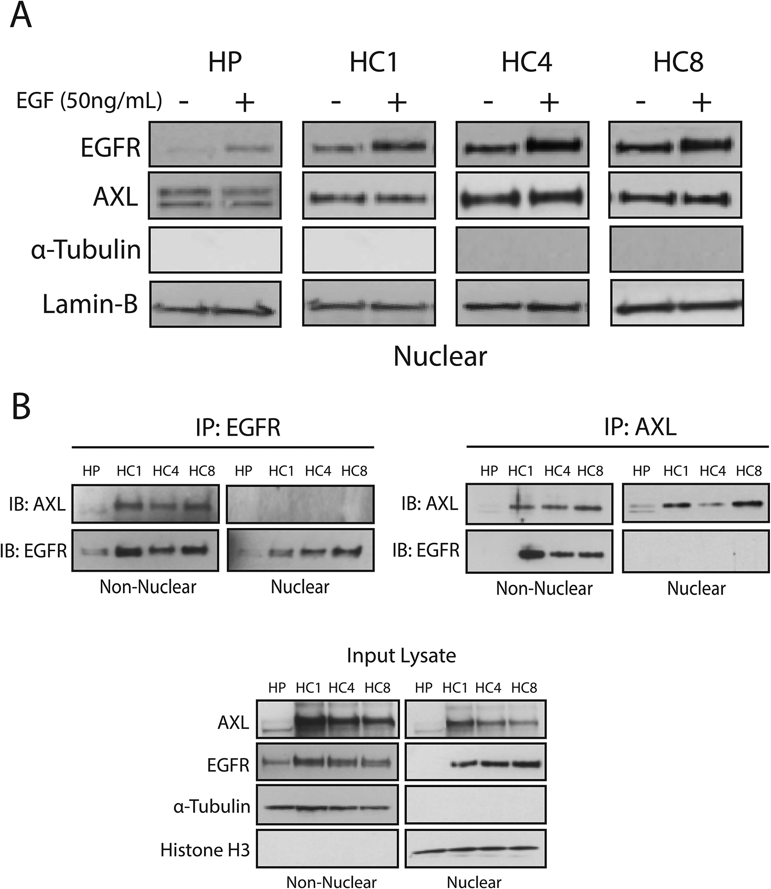
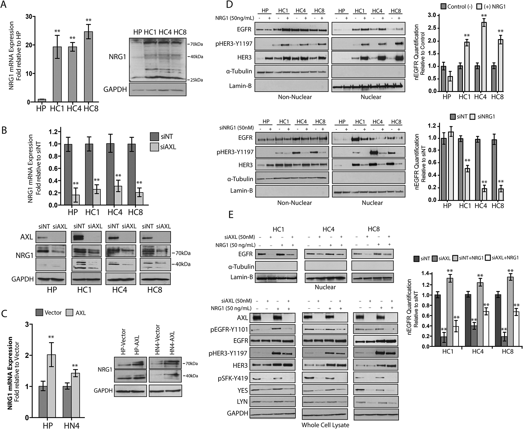
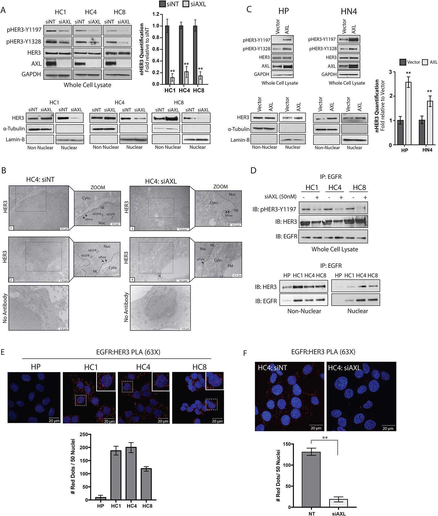
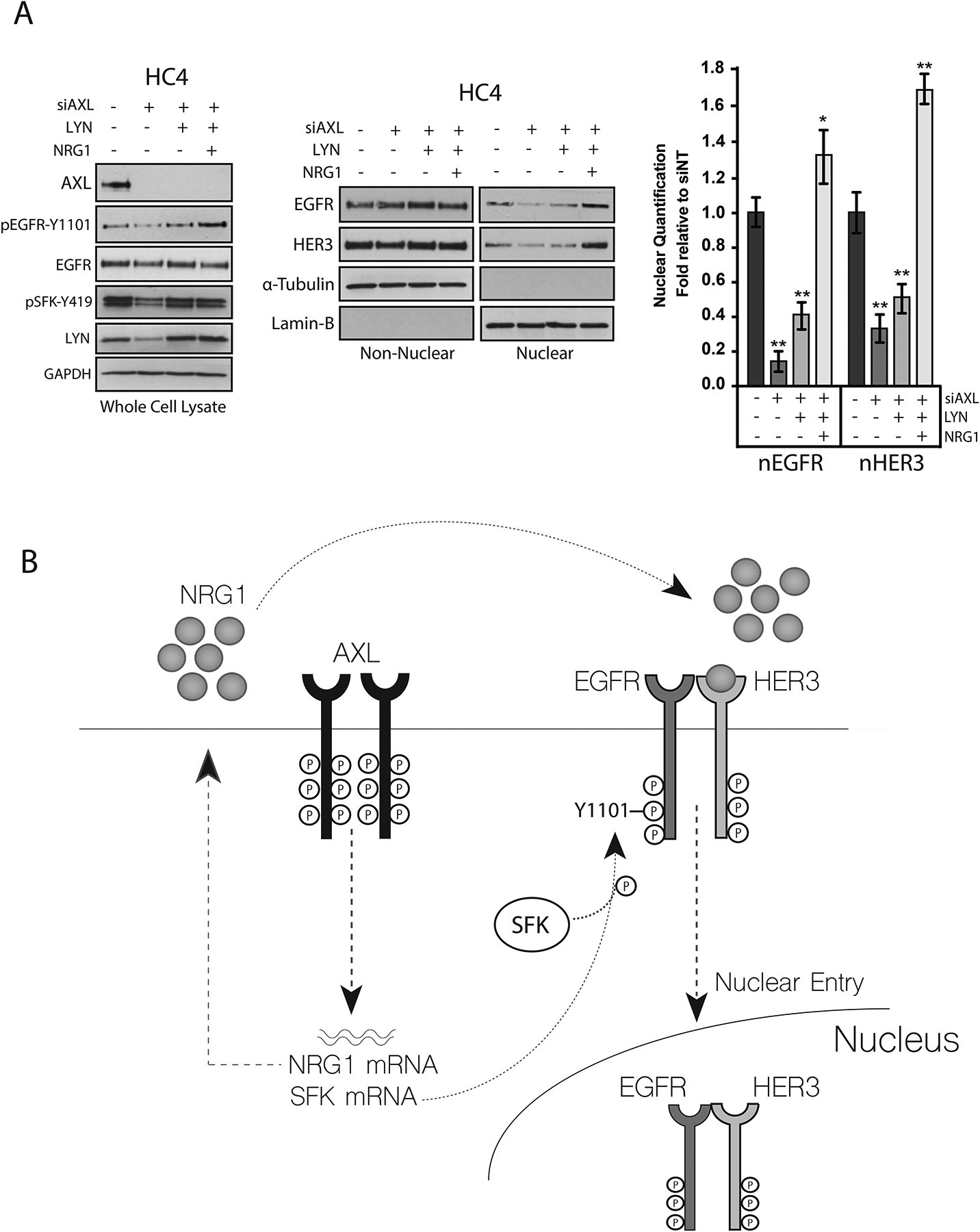
The epidermal growth factor receptor (EGFR) is a therapeutic target in patients with various cancers. Unfortunately, resistance to EGFR-targeted therapeutics is common. Previous studies identified two mechanisms of resistance to the EGFR monoclonal antibody cetuximab. Nuclear translocation of EGFR bypasses the inhibitory effects of cetuximab, and the receptor tyrosine kinase AXL mediates cetuximab resistance by maintaining EGFR activation and downstream signaling. Thus, we hypothesized that AXL mediated the nuclear translocation of EGFR in the setting of cetuximab resistance. Cetuximab-resistant clones of non-small cell lung cancer in culture and patient-derived xenografts in mice had increased abundance of AXL and nuclear EGFR (nEGFR). Cellular fractionation analysis, super-resolution microscopy, and electron microscopy revealed that genetic loss of AXL reduced the accumulation of nEGFR. SRC family kinases (SFKs) and HER family ligands promote the nuclear translocation of EGFR. We found that AXL knockdown reduced the expression of the genes encoding the SFK family members YES and LYN and the ligand neuregulin-1 (NRG1). AXL knockdown also decreased the interaction between EGFR and the related receptor HER3 and accumulation of HER3 in the nucleus. Overexpression of LYN and NRG1 in cells depleted of AXL resulted in accumulation of nEGFR, rescuing the deficit induced by lack of AXL. Collectively, these data uncover a previously unrecognized role for AXL in regulating the nuclear translocation of EGFR and suggest that AXL-mediated SFK and NRG1 expression promote this process.


Conflict of interest statement

Competing interests: No conflicts of interest

Figures

Figure 1:
Figure 1:. CtxR clones and xenografts have increased abundance of nEGFR and AXL.
(A) Immunoblotting (top, left) in non-nuclear and nuclear lysates harvested from three CtxR clones (HC1, HC4, and HC8) and the CtxS parental cell line HP. Calnexin, α-Tubulin, and histone H3 were used as loading and purity controls for non-nuclear and nuclear lysates, respectively. Structured resolution microscopy (bottom) in parental (HP) and CtxR clones stained for DAPI (blue) and EGFR (red) (bottom). Magnification, 100X. Scale bars, 10μm. Transmission electron microscopy (TEM; right) of fixed HC4 cells labeled with EGFR antibody-bound gold particles. Black arrows in insets mark gold particles in the nucleus. Cyto, cytoplasm; NE, nuclear envelope; nPore, nuclear pore; Nuc, nucleus. Scale bars, 0.5 μm. (B) Immunobloting in whole cell lysates from HP and CtxR clones. GAPDH: loading control. (C) Tumor volume (left) and immunohistochemical staining for EGFR and AXL in representative NCI-H226 xenografts (right) from IgG- or cetuximab-treated mice (n=4 and 5 mice, respectively; representative sections from 3 of each group are shown). Magnification, 40X. Scale bars, 1000 μm. (D) Immunohistochemical staining for EGFR and AXL abundance in CtxS and CtxR NSCLC PDXs. Magnification, 20X. Scale bars, 1000 μm. Black arrows in insets mark nEGFR. nEGFR and AXL abundance were quantified with ImageJ software. AXL staining intensity in CtxR tumors was normalized to that of the IgG or CtxS tumors (n=6 mice; representative sections from 3 of each group are shown). Data are means ± SD of 3 independent fields of view per tumor. * P < 0.05; ** P < 0.01, by two-tailed Student t-test. Blots and microscopy are representative of 3 experiments.
Figure 2:
Figure 2:. EGFR nuclear translocation is dependent on AXL.
(A) Non-nuclear and nuclear proteins were harvested from HP and CtxR clones 72 hours after transfection with siAXL or siNT followed by immunoblotting for the indicated proteins. Confocal and super resolution microscopy was performed in HC4 cells 72 hours after transfection with siAXL or siNT. Confocal imaging depicts overlap between DAPI (blue) and EGFR (red). Magnification, 40X. Scale bars, 20 μm. Z-slice imaging depicts this overlap (white dashed-line boxes). Magnification, 60X. Scale bars, 20 μm. Super resolution imaging depicts overlap of DAPI (blue) and EGFR (red). Magnification, 100X. Scale bars, 10 μm. (B) TEM of fixed HC4 cells transfected with siAXL or siNT for 72 hours and subsequently labeled with EGFR antibody-bound gold particles. Black arrows in insets mark gold particles in the nucleus; n= 100 cells analyzed per condition from three independent experiments. Cyto, cytoplasm; NE, nuclear envelope; nPore, nuclear pore; Nuc, nucleus. Scale bars, 0.5 μm (zoom view) or 1.0 μm. (C and D) HC4 cells were transfected with siAXL or siNT for 24 hours prior to overexpression of EGFR-GFP (C) or EGFR-HA (D) for an additional 48 hours. Whole cell, non-nuclear and nuclear lysate was harvested followed by immunoblotting for the indicated proteins. Confocal IF was performed to visualize GFP localization in the nucleus (white arrow). Magnification, 60X. Scale bars, 20 μm. Super resolution microscopy was used to visualize HA localization in the nucleus. Magnification, 100X. Scale bars, 10μm. Inset 1 (C), imunoblotting of non-nuclear and nuclear proteins from HC4 cells 48 hours after transfection with vector, EGFR-WT-GFP or EGFR-Y1101F-GFP. (E) Whole cell, non-nuclear and nuclear proteins were harvested from HP and HN4 cells stably overexpressing pcDNA6.0-AXL or pcDNA6.0-Vector control followed by immunoblotting for the indicated proteins. For presented immunoblots, GAPDH, α-Tubulin, calnexin, and histone-H3 were used as loading and purity controls for whole cell, non-nuclear, and nuclear lysates, respectively. Microscopy and blots are representative of 3 experiments and ImageJ software was used to quantify nEGFR abundance. Data in (A) and (E) are mean ± SD of three independent experiments. ** P < 0.01, by two-tailed Student t-test.
Figure 3:
Figure 3:. AXL mediates EGFR nuclear translocation by enhancing YES and LYN mRNA expression.
(A) HP and CtxR clones were incubated with siAXL or siNT for 72 hours prior to harvesting whole-cell lysate and immunoblotting for the indicated proteins. (B) mRNA was harvested from CtxR clones and HP cells 72 hours post transfection with siAXL or siNT. YES and LYN mRNA expression was detected by qPCR and normalized to the expression of each target in siNT-transfected cells. β-Actin was used as an endogenous control. Data are mean ± SD of 3 in three independent experiments. ** P < 0.01 by the Mann-Whitney U test. (C) Whole cell lysate was harvested from HP and HN4 cells stably overexpressing pcDNA-AXL or Vector control and subsequently subjected to immunoblot analysis. (D) Whole cell, non-nuclear and nuclear proteins were harvested from HP and HN4 cells stimulated with Gas6 (300 ng/mL) for 30 min followed by immunoblotting for the indicated proteins. (E) HC4 cells were transfected with siAXL (50nM) or siNT for 24 hours prior to overexpression of pcDNA6.0-LYN for an additional 48 hours. Whole cell, non-nuclear, and nuclear lysate was subsequently harvested followed by immunoblotting for the indicated proteins. For presented immunoblots, GAPDH, α-Tubulin, and lamin-B were used as loading and purity controls for whole cell, non-nuclear and nuclear lysates, respectively. All immunoblots are representative of three independent experiments, and ImageJ software was used to quantify nEGFR abundance. Data in (E) are mean ± SD of three independent experiments. ** P < 0.01, by two-tailed Student t-tests.
Figure 4:
Figure 4:. EGFR traffics to the nucleus independently from AXL.
(A) HP and CtxR clones were stimulated with EGF (50 ng/mL) for 30 min prior to harvesting nuclear proteins and immunoblot analysis. (B) Non-nuclear and nuclear lysate (500 μg each) harvested from HP and CtxR clones were subjected to immunoprecipitation with an AXL or EGFR antibody. Input lysate was examined for loading and purity controls for the non-nuclear and nuclear fractions, respectively. α-Tubulin, lamin-B and histone H3 were used as loading and purity controls for the nuclear lysates, respectively. Data are representative of three independent experiments.
Figure 5.
Figure 5.. AXL mediates EGFR translocation to the nucleus through NRG1.
(A) NRG1 mRNA expression and protein abundance was detected in HP and CtxR clones. NRG1 mRNA expression was detected by qPCR and normalized to NRG1 expression in HP cells. (B) NRG1 mRNA expression and protein abundance were evaluated in HP and CtxR clones 72 hours post transfection with siAXL (50 nM) or siNT. NRG1 mRNA expression was detected by qPCR and normalized to expression levels detected in cells transfected with siNT. (C) NRG1 mRNA expression and protein abundance were detected in HP-AXL and HN4-AXL stable cell lines. NRG1 mRNA expression was detected by qPCR and normalized to NRG1 expression in HP-Vector or HN4-Vector cells. (D) HP and CtxR clones were stimulated with 50 ng/mL NRG1 for 30 min (top) or transfected with siNRG1 (50 nM) or siNT for 72 hours (bottom) prior to harvesting non-nuclear and nuclear proteins. (E) CtxR clones were transfected with siAXL (50 nM) or siNT for 72 hours prior to stimulation with NRG1 (50 ng/mL) for 30 minutes. Whole cell lysate and nuclear proteins were harvested followed by immunoblot analysis. β-Actin was used as an endogenous control for all qPCR experimentation. Data are mean ± SD of three independent experiments. ** P < 0.01, by the Mann-Whitney U test. For presented immunoblots, GAPDH, α-Tubulin, and lamin-B were used as loading and purity controls for whole cell, non-nuclear and nuclear lysates, respectively. ImageJ software was used to quantify nEGFR abundance. Data are mean ± SD of three independent experiments. ** P < 0.01, by two-tailed Student t-test.
Figure 6.
Figure 6.. AXL stimulates EGFR-HER3 interaction and nuclear translocation.
(A) Whole cell, non-nuclear and nuclear proteins were harvested from CtxR clones 72 hours after transfection with siAXL (50nM) or siNT followed by immunoblotting for the indicated proteins. (B) TEM of fixed HC4 cells transfected with siAXL or siNT for 72 hours and subsequently labeled with HER3 antibody-bound gold particles. Black arrows in insets mark gold particles in the nucleus; n= 100 cells analyzed per condition from three independent experiments. Cyto, cytoplasm; NE, nuclear envelope; nPore, nuclear pore; Nuc, nucleus. Scale bars, 0.2μm, 0.5μm or 1.0μm. (C) Whole cell, non-nuclear, and nuclear proteins were harvested from HP-AXL and HN4-AXL stable cell lines followed by immunoblotting for the indicated proteins. (D) 500 μg of non-nuclear and nuclear lysate harvested from HP and CtxR clones were subjected to IP with an anti-EGFR antibody, followed by immunoblotting for HER3 and EGFR (top). Whole cell lysate was harvested from CtxR clones transfected with siAXL (50 nM) or siNT for 72 hours and examined for EGFR and HER3 interaction via IP with an anti-EGFR antibody (bottom). (E and F) EGFR and HER3 interactions were examined via PLA in CtxR clones and HP cells in (E) and the HC4 clone transfected with siAXL (50nM) or siNT for 72 hours (F). Red dots were counted in 50 nuclei from five-six independent fields of view per cell line. Data are mean ± SD of two independent experiments in (E) and three independent experiments in (F). Magnification, 60X. Scale bars, 20 μm. ** P < 0.01, by two-tailed Student t-test. For presented immunoblots, GAPDH, α-Tubulin, and lamin-B were used as loading and purity controls for whole cell, non-nuclear and nuclear lysates, respectively. ImageJ software was used to quantify nHER3 abundance in (A) and (C). Data are mean ± SD of three independent experiments for all immunoblots. ** P < 0.01, by two-tailed Student t-test.
Figure 7:
Figure 7:. SFKs and NRG1 are necessary and sufficient in mediating EGFR nuclear translocation.
(A) HC4 cells were transfected with siAXL (50nM) or siNT for 24 hours prior to overexpression of pcDNA6.0- LYN for an additional 48 hours. Cells were then stimulated with NRG1 (50 ng/mL) for 30 min followed by harvesting whole cell, non-nuclear, and nuclear proteins. GAPDH, α-Tubulin, and lamin-B were used as loading and purity controls for whole cell, non-nuclear and nuclear lysates, respectively. ImageJ software was used to quantify nEGFR and nHER3 abundance. Data are mean ± SD of three independent experiments. ** P < 0.01, by two-tailed Student t-test. (B) Model of AXL mediated EGFR nuclear translocation that we propose occurs in CtxR cells.

Comment in

  • Findings of Research Misconduct.
    [No authors listed] [No authors listed] Fed Regist. 2022 Apr 11;87(69):21129-21130. Fed Regist. 2022. PMID: 35444354 Free PMC article. No abstract available.

References

    1. Yarden Y, Pines G. The ERBB network: at last, cancer therapy meets systems biology. Nat Rev Cancer. 2012;12(8):553–63. Epub 2012/07/13. doi: 10.1038/nrc3309. - DOI - PubMed
    1. Yarden Y, Shilo BZ. SnapShot: EGFR signaling pathway. Cell. 2007;131(5):1018. doi: 10.1016/j.cell.2007.11.013. - DOI - PubMed
    1. Yewale C, Baradia D, Vhora I, Patil S, Misra A. Epidermal growth factor receptor targeting in cancer: a review of trends and strategies. Biomaterials. 2013;34(34):8690–707. doi: 10.1016/j.biomaterials.2013.07.100. - DOI - PubMed
    1. Han W, Lo HW. Landscape of EGFR signaling network in human cancers: biology and therapeutic response in relation to receptor subcellular locations. Cancer Lett. 2012;318(2):124–34. Epub 2012/01/21. doi: 10.1016/j.canlet.2012.01.011. - DOI - PMC - PubMed
    1. Sorkin A, Goh LK. Endocytosis and intracellular trafficking of ErbBs. Exp Cell Res. 2009;315(4):683–96. - PubMed

Publication types

MeSH terms