. 2017 Jan 17;114(3):E376-E385.
doi: 10.1073/pnas.1619735114. Epub 2017 Jan 3.

Characterization of cytopathic factors through genome-wide analysis of the Zika viral proteins in fission yeast

Affiliations

Characterization of cytopathic factors through genome-wide analysis of the Zika viral proteins in fission yeast

Ge Li et al. Proc Natl Acad Sci U S A. .

Abstract

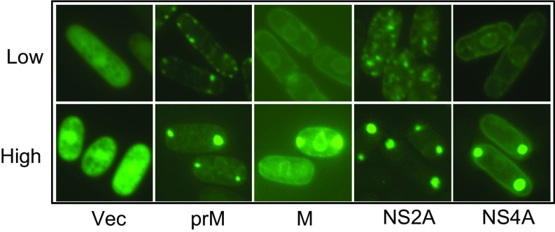
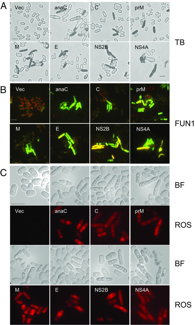
The Zika virus (ZIKV) causes microcephaly and the Guillain-Barré syndrome. Little is known about how ZIKV causes these conditions or which ZIKV viral protein(s) is responsible for the associated ZIKV-induced cytopathic effects, including cell hypertrophy, growth restriction, cell-cycle dysregulation, and cell death. We used fission yeast for the rapid, global functional analysis of the ZIKV genome. All 14 proteins or small peptides were produced under an inducible promoter, and we measured the intracellular localization and the specific effects on ZIKV-associated cytopathic activities of each protein. The subcellular localization of each ZIKV protein was in overall agreement with its predicted protein structure. Five structural and two nonstructural ZIKV proteins showed various levels of cytopathic effects. The expression of these ZIKV proteins restricted cell proliferation, induced hypertrophy, or triggered cellular oxidative stress leading to cell death. The expression of premembrane protein (prM) resulted in cell-cycle G1 accumulation, whereas membrane-anchored capsid (anaC), membrane protein (M), envelope protein (E), and nonstructural protein 4A (NS4A) caused cell-cycle G2/M accumulation. A mechanistic study revealed that NS4A-induced cellular hypertrophy and growth restriction were mediated specifically through the target of rapamycin (TOR) cellular stress pathway involving Tor1 and type 2A phosphatase activator Tip41. These findings should provide a reference for future research on the prevention and treatment of ZIKV diseases.

Keywords: Schizosaccharomyces pombe; Zika genome; Zika proteins; cytopathic factors; fission yeast.


Conflict of interest statement

The authors declare no conflict of interest.

Figures

Fig. 1.
Fig. 1.
Genome-wide analysis of intracellular localizations of ZIKV proteins. (A) Schematic structure of the ZIKV genome with the predicted association of each ZIKV protein with the intracellular membrane. The ZIKV genome shown is based on the MR766 viral genome (GenBank accession number: Ay632535). Each of the viral proteins is drawn based on the relative orientation in the entire RNA genome. The ZIKV viral protease, host protease, and Furin protease are indicated by arrows. Each arrow points to the specific protease cleavage site. The numbers shown above each protein product indicate the start/end position. NS5 encodes methyltransferase at its N-terminal end and RNA-dependent RNA polymerase at its C-terminal end. ZIKV protein products information based on Kuno et al. (16). The protein membrane-associated structure and processing are information from Assenberg et al. (17). (B) The primary intracellular localizations of ZIKV proteins. All GFP-ZIKV proteins were produced at low gene-expression levels and observed within 20 h of GI. (a) An empty vector control (Vec). (b) Structural ZIKV proteins. (c) Nonstructural ZIKV proteins. (Scale bars, 10 μm.) (C, a) Comparison of ZIKV protein localization with cellular proteins that are known to localize in ER (Gpi1-YFP), Golgi (Ynd1-YFP), and cytoplasmic puncta (Atg1-YFP) (1). (b) Colocalization of GFP-NS1 with Atg1-YFP under nitrogen starvation. Note that both GFP and YFP were detectable under the GFP/YFP filter. However, only the GFP signal was detectable under the CFP filter. Arrows indicate where Atg1-YFP signals are missing from the CFP filter. (Scale bar, 10 μm.) The interpretation of the specific subcellular localization of each ZIKV protein is summarized in Table 1. A comparison of subcellular localization patterns of each ZIKV protein at low and high expression levels is presented in Fig. S2.
Fig. S1.
Fig. S1.
Confirmation of molecular cloning of the ZIKV MR766 viral genome into the fission yeast gene-expression pYZ1N and pYZ3N system. (A) Molecular cloning into the pYZ1N gene expression vector. (a) Results of PCR-amplified products of each ZIKV ORF-encoding DNA sequence from ZIKV viral cDNA. (b) Endonuclease restriction digestions of the ZIKV ORF-encoding DNA inserts that were cloned into the pYZ1N. Plasmid DNAs were propagated and isolated from E. coli. (c) Results of yeast colony PCR showing correct inserts of respective ZIKV ORF-carrying plasmids from transformed fission yeast SP223 cells. (B, ac) Gels for molecular cloning of the ZIKV genome into the pYZ3N vector are shown as in A. The order of ORFs from 1–14: anaC, C, PR, M, prM, E, 2K, NS1, NS2A, NS2B, NS3, NS4A, NS4B, and NS5. M, molecular marker.
Fig. S2.
Fig. S2.
Comparison of intracellular localization patterns of ZIKV proteins under high and low levels of protein expression. Only ZIKV proteins that showed differences in localization patterns and possible cytoplasmic puncta are shown here. All GFP-ZIKV proteins were expressed from the pYZ3N gene-expression vector through the inducible nmt1 promoter. The high level of ZIKV protein was produced with full induction of the nmt1 promoter by complete removal of thiamine from the growth medium. The subcellular localizations of each ZIKV protein were observed between 24 and 48 h after GI. The low level of ZIKV protein was produced with partial induction of the nmt1 promoter using 10 nM thiamine in the growth medium (14, 51). The subcellular localizations of each ZIKV protein were observed within 20 h after GI.
Fig. 2.
Fig. 2.
The cytopathic effects of ZIKV proteins. (A) The effects on cell proliferation. (a) Expression of ZIKV mRNA transcripts measured 24 h after GI by RT-PCR. The order of proteins from 1–14: anaC, C, PR, M, prM, E, 2K, NS1, NS2A, NS2B, NS3, NS4A, NS4B, and NS5. M, molecular marker. (b) Effect of ZIKV expression on fission yeast colony formation. Agar plates are numbered as in a. An empty pYZ1N vector was used as a control and designated as zero. ZIKV-carrying cells grown on selective EMM agar plates were incubated at 30 °C for 4–5 d before the images were captured. Off, gene-suppressed; On, gene-induced. (c) Growth curve analysis was used to quantify growth inhibition by ZIKV proteins. Only the effect of ZIKV proteins (anaC, C, E, prM, M, NS2B, and NS4A) that affected yeast colony-forming abilities as demonstrated in b are shown. Cell growth was measured spectrophotometrically at OD650 over the indicated time period. The experiment was repeated at least three times, and the SEs of each time point were calculated. ●, gene-on, the ZIKV gene was induced; ○, gene-off, the ZIKV gene was suppressed. (B) The effects on cell morphology. Only ZIKV proteins that affected cell proliferation as demonstrated in A, c are shown. The effects of all ZIKV proteins on fission yeast nuclear morphology are included in Fig. S3. (a) Individual cell morphology. Each image was taken 45 h after GI using bright-field microscopy. Note cell hypertrophy (arrow) in the NS4A-expressing cells. Vec, empty vector. (b) Overall cell morphology as shown by the FSC analysis. Ten thousand cells were measured 48 h after GI. The FSC measures the distribution of all cell sizes. The SSC determines intracellular complexity. (C) Effects on nuclear morphology and cell-cycle regulation. (a) Nuclear morphology. Cells were stained with Hoechst blue fluorescent DNA dye. All cell and nuclear morphologies were examined 45 h after GI. (Scale bar, 10 μm.) Effects of all ZIKV proteins on fission yeast nuclear morphology are included in Fig. S4. (b) The effect of ZIKV protein on cell-cycle G1 regulation. Cells were grown in regular EMM medium in which cells normally reside in the G2/M phase of the cell cycle. (c) Effect of ZIKV proteins on cell-cycle G2/M regulation. Cells were grown in a low-nitrogen EMM medium that enriches cells in the G1 phase of the cell cycle, as described previously (1, 2). Cell-cycle profiles were measured by DNA content using flow cytometric analysis 48 h after GI. Average and SD values were calculated based on the results of three independent experiments. A pairwise Student t test was conducted to compare the DNA content values of each ZIKV protein with and without GI. *P < 0.01. Arrows indicate the location of differences (C, b and c) or where the abnormal cells reside (B, b and C, a).
Fig. S3.
Fig. S3.
Effect of ZIKV protein production on fission yeast cell morphology. (A) Individual cell morphology. (a) Structural ZIKV proteins. (b) Nonstructural ZIKV proteins. Each image was obtained 45 h after GI using bright-field microscopy. (B) Overall cell morphology as shown by FSC analysis. Ten thousands cells were measured 48 h after GI. The FSC measures the distribution of all cell sizes. The SSC determines intracellular complexity. (a) Structural ZIKV proteins. (b) Nonstructural ZIKV proteins.
Fig. S4.
Fig. S4.
Effects of ZIKV proteins on nuclear morphology and cell-cycle regulation. (A) Nuclear morphology. Cells were stained with Hoechst blue fluorescent DNA dye. All cell and nuclear morphologies were examined 45 h after GI. (a) An empty pYZ1N vector control. (b) Structural ZIKV proteins. (c) Nonstructural ZIKV proteins. (B) Cell-cycle profiling with starting cells predominantly in the G2 phase of the cell cycle. All cells were grown in regular EMM medium. The effect of ZIKV protein on the cell cycle was measured by flow cytometry 48 h after GI. (a) Structural ZIKV proteins. (b) Nonstructural ZIKV proteins. (C) Cell-cycle profiling with starting cells predominantly in the G1 phase of the cell cycle. All cells were grown in the low-nitrogen EMM medium to enrich G1 cells (6). The effect of ZIKV protein on cell cycle was measured by flow cytometry 48 h after GI. (a) Structural ZIKV proteins. (b) Nonstructural ZIKV proteins.
Fig. 3.
Fig. 3.
ZIKV proteins induce cell death and cellular oxidative stress. ZIKV-induced cell death was measured 48 h after GI by Trypan blue (TB) staining (A) and the yeast live/dead assay (B), which was measured by FUN-1 staining 40–45 h after GI. (C) ZIKV induces oxidative stress, as indicated by DHE staining showing ROS expression. Images were taken 48 h after GI. (Scale bar, 10 μm.) BF, bright-field.
Fig. 4.
Fig. 4.
NS4A impacts the TOR pathway. (A) The effect of TOR pathway-related gene deletions on NS4A-induced hypertrophy and cellular growth. (a) Cell morphologic changes when NS4A protein was expressed 48 h after GI in wild-type, ∆Tor1, and ∆Tip41 mutant strains. Note that NS4A induced hypertrophy in wild-type cells. However, no apparent gross cell enlargement was seen in the NS4A-expressing ∆Tor1 cells. Conversely, NS4A induced a spherical cell phenotype in the ∆Tip41 mutant strain. (b) Overexpression of the Tip41 gene in the ∆Tip41 mutant strain produced a spherical cell phenotype similar to that shown in a. (Scale bar, 10 μm.) (B) ZIKV NS4A gene transcription by RT-PCR 24 h after GI. (C) The ∆Tor1 deletion suppressed the effect of NS4A on yeast colony formation. (D) ∆Tor1 deletion restored (↑) cellular growth to nearly the normal level in the NS4A-expressing cells; ∆Tip41 worsened NS4A-induced growth inhibition (↓). ●, NS4A gene expressed; ○, NS4A gene suppressed.

References

    1. Dang J, et al. Zika virus depletes neural progenitors in human cerebral organoids through activation of the innate immune receptor TLR3. Cell Stem Cell. 2016;19(2):258–265. - PMC - PubMed
    1. Qian X, et al. Brain-region-specific organoids using mini-bioreactors for modeling ZIKV exposure. Cell. 2016;165(5):1238–1254. - PMC - PubMed
    1. Cugola FR, et al. The Brazilian Zika virus strain causes birth defects in experimental models. Nature. 2016;534(7606):267–271. - PMC - PubMed
    1. Cao-Lormeau VM, et al. Guillain-Barré syndrome outbreak associated with Zika virus infection in French Polynesia: A case-control study. Lancet. 2016;387(10027):1531–1539. - PMC - PubMed
    1. Smith DW, Mackenzie J. Zika virus and Guillain-Barré syndrome: Another viral cause to add to the list. Lancet. 2016;387(10027):1486–1488. - PubMed

Publication types

MeSH terms

Substances