. 2017 Jan 17;114(3):580-585.
doi: 10.1073/pnas.1614035114. Epub 2017 Jan 3.

Gpr132 sensing of lactate mediates tumor-macrophage interplay to promote breast cancer metastasis

Affiliations

Gpr132 sensing of lactate mediates tumor-macrophage interplay to promote breast cancer metastasis

Peiwen Chen et al. Proc Natl Acad Sci U S A. .

Abstract

Macrophages are prominent immune cells in the tumor microenvironment that exert potent effects on cancer metastasis. However, the signals and receivers for the tumor-macrophage communication remain enigmatic. Here, we show that G protein-coupled receptor 132 (Gpr132) functions as a key macrophage sensor of the rising lactate in the acidic tumor milieu to mediate the reciprocal interaction between cancer cells and macrophages during breast cancer metastasis. Lactate activates macrophage Gpr132 to promote the alternatively activated macrophage (M2)-like phenotype, which, in turn, facilitates cancer cell adhesion, migration, and invasion. Consequently, Gpr132 deletion reduces M2 macrophages and impedes breast cancer lung metastasis in mice. Clinically, Gpr132 expression positively correlates with M2 macrophages, metastasis, and poor prognosis in patients with breast cancer. These findings uncover the lactate-Gpr132 axis as a driver of breast cancer metastasis by stimulating tumor-macrophage interplay, and reveal potential new therapeutic targets for breast cancer treatment.

Keywords: Gpr132; breast cancer; lactate; macrophage; metastasis.


Conflict of interest statement

The authors declare no conflict of interest.

Figures
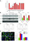
Fig. 1.
Fig. 1.
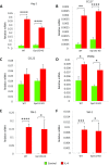
Tumor-derived factors activate M2-like macrophages via Gpr132. (A) pH values of CM from cancer cells or macrophages (n = 5–8). *P < 0.05, ****P < 0.001 compared with RAW 264.7 macrophage CM. (B) Western blot for CD206 and Gpr132 in RAW 264.7 macrophages after treatment with the indicated cancer cell CM for 24 h. Actin was used as a loading control. The CD206/actin or Gpr132/actin ratio was quantified and is shown as fold changes compared with control (n = 3). *P < 0.05, ***P < 0.005 compared with control. (C) RT-qPCR analysis of Gpr132 and CD206 mRNA in BMDMs with or without EO771 CM treatment (n = 3). *P < 0.05. (D) Flow cytometry analysis of Gpr132 and CD206 in BMDMs with or without EO771 CM treatment. The experiments were repeated twice, and the representative results are shown. (E) Immunofluorescence staining for CD11b in WT and Gpr132-KO BMDMs after differentiation in the absence or presence of 30% (vol/vol) EO771 CM or 4T1.2 CM for 7 d. Elongated macrophage morphology indicates an M2-like phenotype. Nuclei were stained with DAPI. (Left) Representative images. (Scale bar, 25 μm.) (Right) Quantification of macrophage morphology as an elongation factor (n = 2–4). ****P < 0.001.
Fig. S1.
Fig. S1.
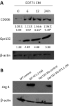
Cancer cell-derived factors promote macrophage M2 activation via Gpr132. (A) Western blot for CD206 and Gpr132 in RAW 264.7 macrophages after treatment with EO771 CM for 0–24 h. Actin was used as a loading control. The CD206/actin or Gpr132/actin ratio was quantified and is shown as fold changes compared with control (0 h). *P < 0.05 compared with control (0 h). (B) Western blot for Arg-1 in WT and Gpr132-KO BMDMs after treatment with 4T1.2 CM for 24 h. Actin was used as a loading control.
Fig. S2.
Fig. S2.
Less than a 3-kDa fraction of cancer cell CM displays lower pH and induces the macrophage M2 phenotype. (A) pH values in the whole media and <3-kDa fraction or >3-kDa fraction of RPMI-1640 CM or EO771 CM (n = 3–4). ****P < 0.001. (B) Western blot for CD206 in BMDMs after treatment with whole media and <3-kDa fraction or >3-kDa fraction of RPMI-1640 CM for 24 h. Actin was used as a loading control. The CD206/actin ratio was quantified and is shown as fold changes compared with the group treated with whole media. (C) Western blot for CD206 in WT and Gpr132-KO BMDMs after treatment with mock control, the whole CM, and the <3-kDa fraction or >3-kDa fraction of CM for 24 h. Actin was used as a loading control. The CD206/actin ratio was quantified and is shown as fold changes compared with WT BMDMs treated with mock control.
Fig. S3.
Fig. S3.
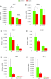
Cancer cell-derived lipids reduce macrophage M2 phenotype independent of Gpr132. RT-qPCR analysis of the expression of Arg-1 (A), PPAR-γ (B), CCL17 (C), CCL22 (D), YM-1 (E) and YM-2 (F) in WT or Gpr132-KO spleen macrophages treated with cancer cell-derived lipids or vehicle control for 24 h (n = 3). *P < 0.05; ***P < 0.005.
Fig. 2.
Fig. 2.
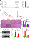
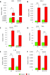
Lactate is a Gpr132 ligand/activator that induces the macrophage M2 phenotype. (A) Lactate in the EO771 <3-kDa CM bound to macrophage Gpr132. Liquid chromatography-mass spectrometry (LC-MS) was used to quantify lactate in the eluent of Gpr132 coimmunoprecipitation (IP). (Left) Representative LC-MS chromatograms. MA, integrated peak mass area (area under curve); RT, retention time. (Right) Quantification of relative lactate binding (n = 3). ****P < 0.001 compared with WT. (B) Calcium mobilization triggered by lactate, but not HCl, was significantly impaired in Gpr132-KO BMDMs. WT or Gpr132-KO BMDMs were stimulated with 25 mM lactate (Left) or HCl (Right) (n = 3). (C) Macrophage morphology after cancer cell CM treatment. The 4T1.2 cells were cultured in the presence or absence of oxamic acid (90 mM) for 3 d. CM was harvested after the cells were cultured for another 24 h without oxamic acid. As a rescue treatment, CM of oxamic acid-treated 4T1.2 cells was supplemented with exogenous lactate. (Left) Representative images of WT and Gpr132-KO BMDMs treated with the indicated CM for 24 h. (Scale bar, 500 μm.) (Right) Quantification of macrophage morphology as an elongation factor (n = 2–3). *P < 0.05, ***P < 0.005. (D) Western blot for CD206 in WT and Gpr132-KO BMDMs after treatment with lactate (5 mM) for 24 h. Actin was used as a loading control. The CD206/actin ratio was quantified and is shown as fold changes compared with control (n = 4). RT-qPCR analysis of the expression of GM-CSF (E) and CCL17 (F) in WT or Gpr132-KO BMDMs in the presence or absence of lactate (25 mM) for 6 h (n = 3–4). In DF, *P < 0.05 compared with vehicle control in the same genotype, ##P < 0.05 and #P < 0.1 compared with WT control under the same treatment condition.
Fig. S4.
Fig. S4.
Lactate secreted by EO771 and 4T1.2 cells is responsible for the low pH in their CM. (A) Lactate levels in the CM from various cancer cells (n = 3) (*P < 0.05, ****P < 0.001) compared with EO771 CM (#### P < 0.001) compared with 4T1.2 CM. (B) Lactate levels in EO771 CM at the indicated time point of culture (n = 3). (C) Lactate levels in the whole CM and <3-kDa and >3-kDa fractions of CM from 24-h EO771 cell cultures (n = 3). (D and E) EO771 and 4T1.2 cells were treated with or without oxamic acid (90 mM) for 3 d. CM was harvested after the cells were cultured for another 24 h without oxamic acid before lactate (D) and pH (E) quantification (n = 3). **P < 0.01, ***P < 0.005, ****P < 0.001.
Fig. S5.
Fig. S5.
Lactate activates macrophage M2 phenotype via Gpr132. (A) Macrophage morphology after cancer cell CM treatment. EO771 cells were cultured in the presence or absence of oxamic acid (90 mM) for 3 d. CM was harvested after the cells were cultured for another 24 h without oxamic acid. Representative images are shown for WT and Gpr132-KO BMDMs treated with the indicated CM for 24 h. (Scale bar, 500 μm.) The macrophage morphology was shown as an elongation factor by quantification of the length of the long axis divided by length of the short axis (n = 2–3). **P < 0.01, ***P < 0.005. (B) Western blot for CD206 in WT and Gpr132-KO spleen macrophages after treatment with lactate (5 mM) for 24 h. Actin was used as a loading control.
Fig. S6.
Fig. S6.
Gpr132 is not required for IL-4 induction of M2 macrophage. RT-qPCR analysis of the expression of Arg-1 (A), CCL17 (B), CCL22 (C), PPAR-γ (D), YM-1 (E), and YM-2 (F) in WT or Gpr132-KO spleen macrophages treated with IL-4 (10 ng/mL) or vehicle control (n = 3) for 16 h. *P < 0.05, **P < 0.01, ***P < 0.005, ****P < 0.001.
Fig. S7.
Fig. S7.
Gpr132 is not required for lipopolysaccharide (LPS) induction of M1 macrophages. RT-qPCR analysis of the expression of inducible nitric oxide synthase (iNOS) (A), COX-2 (B), monocyte chemoattractant protein 1 (MCP-1) (C), TNF-α (D), IL-1β (E), and IL-6 (F) in WT or Gpr132-KO BMDMs treated with LPS (50 ng/mL) or vehicle control for 16 h (n = 3–4). *P < 0.05, **P < 0.01, ***P < 0.005, ****P < 0.001.
Fig. 3.
Fig. 3.
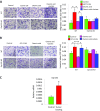
Lactate-activated macrophage promotes cancer cell adhesion, migration, and invasion via Gpr132. (A) Adherence assays. The 4T1.2 cells were suspended in fibronectin (10 mg/mL)-precoated plates with CM from spleen-derived macrophages (mf) that were treated with 4T1.2 CM or lactate (5 mM) with or without Gpr132 antibody (6 mg/mL) or normal IgG (6 mg/mL). The adhered cells were stained with crystal violet, dissolved in 1% Triton X-100, and measured at OD590. (B and C) Boyden chamber assay of cancer cell migration and invasion. The 4T1.2 cells (B) or EO771 cells (C) were plated on the upper chamber inserts with untreated (Control), and 4T1.2 CM-activated or lactate-activated spleen-derived WT or Gpr132-KO macrophages plated in the lower chambers. For the invasion assay, the inserts were precoated with 60 μL of Matrigel. After migration for 6 h (B) or invasion for 24 h (C), the migrated or invaded cells were stained with crystal violet and counted as cells per field of view under the microscope (n = 3–4). *P < 0.05, **P < 0.01, ***P < 0.005, ****P < 0.001. (Scale bars, 500 μm.)
Fig. S8.
Fig. S8.
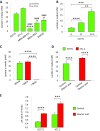
Cancer cell-derived lactate activates macrophages to promote cancer cell migration and invasion via Gpr132. (A and B) Boyden chamber assay. EO771 or 4T1.2 cells were cultured in the presence or absence of oxamic acid (90 mM) for 3 d. CM was harvested after the cells were cultured for another 24 h without oxamic acid. As a rescue, CM of oxamic acid-treated EO771 or 4T1.2 cells was supplemented with exogenous lactate. CM was used to treat WT or Gpr132-KO BMDMs plated in the lower chambers before EO771 cells (A) or 4T1.2 cells (B) were plated on the upper cell culture inserts. For invasion assay, the inserts were precoated with 60 μL of Matrigel. After migration for 6 h (A) or invasion for 24 h (B), the migrated or invaded cells were stained with crystal violet and counted as cells per field of view under the microscope (n = 3–4). *P < 0.05, ****P < 0.001. (Scale bars, 500 μm.) (C) Primary breast tumor enhances Gpr132 expression in distant metastatic sites. EO771 cells were transplanted into the mammary fat pad of WT mice. When the primary tumors reached about 1,500 mm3, lungs were harvested. Gpr132 expression in the lung of control and tumor-bearing mice was quantified by RT-qPCR (n = 3–4). *P < 0.05 compared with control.
Fig. 4.
Fig. 4.
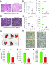
Gpr132 deletion attenuates breast cancer lung metastasis by reducing M2 macrophages. (AG) Lung metastases from breast cancer cells were decreased in Gpr132-KO mice. EO771 cells (AC) or EO771-LMB cells (DG) were transplanted into the mammary fat pad of WT and Gpr132-KO mice. Primary tumors were resected when they reached 1,500 mm3 (AC) or 650 mm3 (DG). After 25 d (AC) or 16 d (DG), lungs were harvested and subjected to H&E staining (A and D). Arrows indicate metastatic foci. The number (B and E) and size (C and F) of tumor nodules were quantified from the stained lung sections (n = 7–9). (G) Lung metastases were quantified using a fluorescent probe that selectively activates in tumors, but not normal tissues, by responding to low pH. In the EO771-LMB model, mice were injected i.v. with Probe 5c 24 h before lung dissection and image acquisition (n = 2–3). (HK) M2 macrophages in lung metastasis were reduced in Gpr132-KO mice. IHC was performed for CD206 (H), as well as RT-qPCR for Arg-1 (I), GM-CSF (J), and CCL22 (K), in the lungs of WT or Gpr132-KO mice of the EO771-LMB model (n = 4). *P < 0.05, ***P < 0.005. (Scale bars, 500 μm.)
Fig. S9.
Fig. S9.
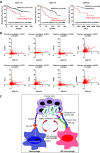
Gpr132 expression positively correlates with metastasis and M2 macrophage in human breast cancer. (A) Higher Gpr132 expression correlated with lower metastasis-free and relapse-free survival in patients with breast cancer. Data were obtained from PrognoScan. (B) Linear regression analyses of TCGA-BRCA RNA-seq data showed that Gpr132 expression positively correlated with the expression of M2 macrophage markers, including CD163, CCL17, CCL22, C-C chemokine receptor type 2 (CCR2), TLR1, TLR8, transglutaminase 2 (TGM2), and CD200R1, in human breast cancer lesions (n = 1,100). (C) Working model for how the lactate-Gpr132 axis facilitates a positive feedback loop between the tumor and macrophages to promote breast cancer metastasis. Cancer cell-secreted lactate activates macrophage Gpr132 receptor to induce M2 polarization. In turn, lactate-activated macrophages facilitate breast cancer metastasis. Thus, Gpr132 functions as a macrophage sensor to relay the acidic signal in the tumor microenvironment to TAM activation and metastasis induction.

References

    1. Acharyya S, et al. A CXCL1 paracrine network links cancer chemoresistance and metastasis. Cell. 2012;150(1):165–178. - PMC - PubMed
    1. Jones SE. Metastatic breast cancer: The treatment challenge. Clin Breast Cancer. 2008;8(3):224–233. - PubMed
    1. Condeelis J, Pollard JW. Macrophages: Obligate partners for tumor cell migration, invasion, and metastasis. Cell. 2006;124(2):263–266. - PubMed
    1. Khamis ZI, Sahab ZJ, Sang QX. Active roles of tumor stroma in breast cancer metastasis. Int J Breast Cancer. 2012;2012:574025. - PMC - PubMed
    1. Mao Y, Keller ET, Garfield DH, Shen K, Wang J. Stromal cells in tumor microenvironment and breast cancer. Cancer Metastasis Rev. 2013;32(1-2):303–315. - PMC - PubMed

Publication types

MeSH terms