. 2016 Aug:114:T7.

The Hematologic Definition of Monoclonal Gammopathy of Undetermined Significance in Relation to Paraproteinemic Keratopathy (An American Ophthalmological Society Thesis)

Affiliations

The Hematologic Definition of Monoclonal Gammopathy of Undetermined Significance in Relation to Paraproteinemic Keratopathy (An American Ophthalmological Society Thesis)

Walter Lisch et al. Trans Am Ophthalmol Soc. 2016 Aug.

Erratum in

Abstract

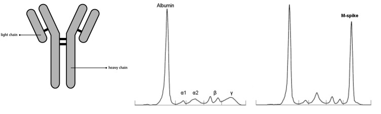
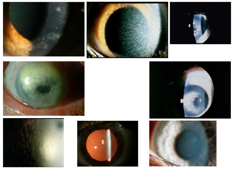
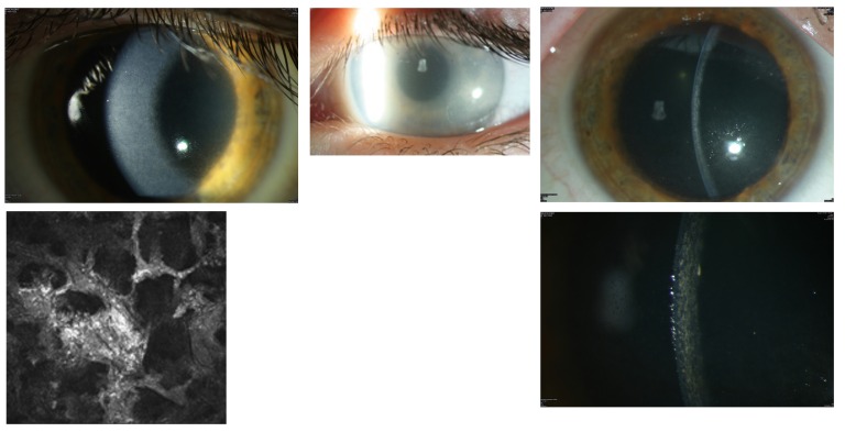
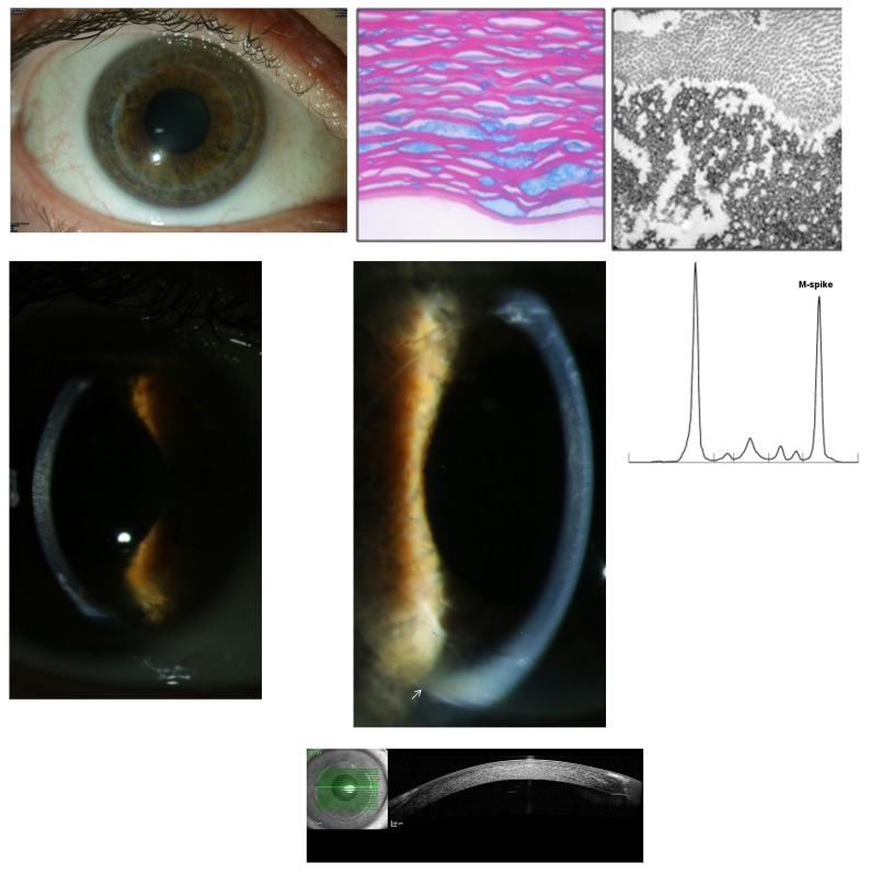
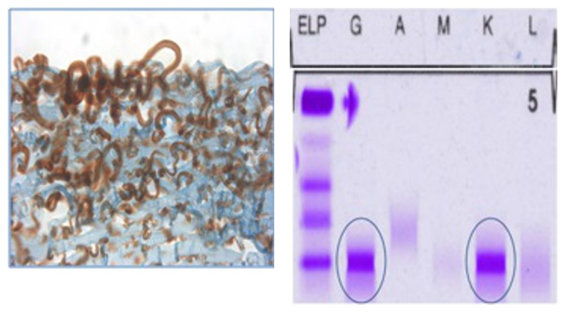
Purpose: To determine if paraproteinemic keratopathy (PPK) in the setting of monoclonal gammopathy of undetermined significance (MGUS) causes distinct patterns of corneal opacification that can be distinguished from hereditary, immunologic, or inflammatory causes.

Methods: A retrospective, interventional study of patients showed distinct bilateral opacity patterns of the cornea at the eye clinics of Hanau, Mainz, Helsinki, Marburg, and Berlin between 1993 and 2015. Data on patient characteristics and clinical features on ophthalmic examination were collected, and serum protein profiles were evaluated. A literature review and analysis of all published studies of MGUS with PPK is also presented.

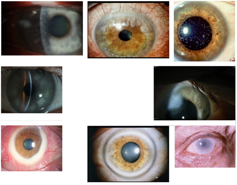
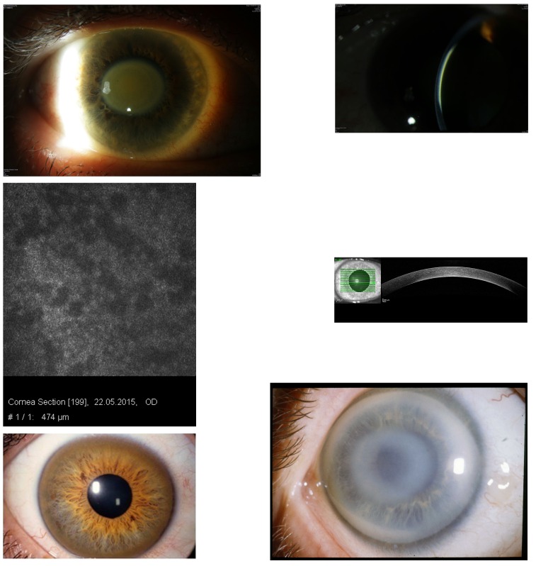
Results: The largest group of patients diagnosed with MGUS-induced PPK is analyzed in this study. We studied 22 eyes of 11 patients (6 male, aged 43 to 65, mean age 54; 5 female, aged 49 to 76, mean age 61) with distinct corneal opacities and visual impairment who were first suspected of having hereditary, inflammatory, or immunologic corneal entities. Subsequently, serum protein electrophoresis revealed MGUS to be the cause of the PPK. Literature review revealed 72 patients with bilateral PPK (34 male, mean age 57; 38 female, mean age 58) in 51 studies of MGUS published from 1934 to 2015 and disclosed six additional corneal opacity patterns.

Conclusions: This thesis shows that MGUS is not always an asymptomatic disorder, in contrast to the hematologic definition, which has no hint of PPK. The MGUS-induced PPK can mimic many other diseases of the anterior layer of the eye. A new clinical classification for PPK in MGUS is proposed.


Figures

FIGURE 1
FIGURE 1
Left, Structure of an immunoglobulin, composed of four polypeptide chains: two light chains (λ and κ) and two heavy chains. Middle, Normal pattern of serum protein electrophoresis. Right, Abnormal pattern of serum protein electrophoresis in a patient with monoclonal gammopathy presenting as the large M spike in the γ region.
FIGURE 2
FIGURE 2
Top row, Case 1 Table 2, MGUS-induced punctiform corneal crystals + patches (left) vs punctiform crystals in cystinosis without patches (middle) and vs crowded comma-shaped crystals of the crystalline type of Schnyder corneal dystrophy + haze (right). Middle row, Case 2 Table 2, MGUS-induced anterior comma-shaped crystals (left) vs crowded comma-shaped crystals of Schnyder corneal dystrophy (right). Bottom row, Case 3 Table 2, MGUS-induced lattice-like lines in retroillumination from the iris (left) vs true lattice lines in retroillumination from the retina of lattice corneal dystrophy type 1 (middle); disappearing of the lattice lines of case 3 and transforming of mild diffuse opacities with focal epithelium elevations after a follow-up of 22 years (right).
FIGURE 3
FIGURE 3
Top row, Case 4 Table 2, MGUS-induced central granules (left) and Case 5 Table 2, MGUS-induced peripheral granules (middle) vs granules of granular corneal dystrophy type 1 (right). Middle row, Case 6 Table 2, MGUS-induced peripheral patch-like opacities (left) vs peripheral hypertrophic degeneration (right). Bottom row, Case 7 Table 2, MGUS-induced peripheral large circular yellowish band with conjunctival injection (left) vs arcus lipoides (middle) and vs peripheral ring of lecithin cholesterol acetyltransferase deficiency (right).
FIGURE 4
FIGURE 4
Case 8 Table 2, MGUS-induced superficial geographic-like opacity with transparent limbus zone (left) vs geographic-like opacity pattern of Reis-Bücklers corneal dystrophy (right).
FIGURE 5
FIGURE 5
Case 10 Table 2, central discoid, golden-brown discoloration of the cornea in a 59-year-old female patient with biclonal gammopathy of undetermined significance and hypercupremia (top left). The thin slit beam shows that the opacity is at the level of Descemet’s membrane (top right). Confocal microscopy at the level of 474 μm reveals irregular dark areas, which partly cover the endothelial cells (middle left). Optical coherence tomography shows the opacity on the posterior cornea of the patient (middle right). Kayser-Fleischer peripheral brownish ring in Wilson’s disease (bottom left). Noncrystalline Schnyder corneal dystrophy with subepithelial central opacity and arcus lipoides (bottom right).
FIGURE 6
FIGURE 6
Case 11 Table 2, a 43-year-old male patient with MGUS-induced flake-like corneal opacification in 2009 (top left) vs flake-like phenotype of congenital stromal corneal dystrophy (top middle). Thin slit beam shows that the MGUS opacity involves the whole stroma (top right). Confocal microscopy shows large bands that mask the keratocytes (bottom left) and progression of the stromal opacities on the right eye in 2012 (bottom right).
FIGURE 7
FIGURE 7
Case 11 Table 2. Top row, Left eye 12 month after penetrating keratoplasty showing no complications (left); histologic evaluation of penetrating keratoplasty specimens demonstrates irregular corneal deposits exhibiting placoid configuration (Masson’s trichrome, ×136) (middle); and transmission electron microscopy image demonstrating stromal extracellular, electron-dense deposits and on the superior part of the image normal collagen fibrils (×81,000) (right). Middle row, Recurrence of stromal involvement, identical to the primary opacity pattern, in the graft of the right eye (left) and of the left eye (arrow localizing graft’s border) in 2014 (middle); protein electrophoresis of the serum with an elevated M spike (right). Bottom, OCT of the right graft showing dense stromal opacities.
FIGURE 8
FIGURE 8
Case 11 Table 2, Immunhistochemistry of the cornea in 2015 (left) showing brownish staining of subepithelial material in an immunoperoxidase reaction for IgG, and with agarose gel electrophoresis of the serum in 2015 (right) that confirms an MGUS-induced paraproteinemic keratopathy of IgGκ light chain type, identified with blue circles; ELP, electrophoresis; G, IgG; A, IgA; M, IgM; K, kappa light chain; L, lambda light chain.

References

    1. Crawford J, Eye MK, Cohen HJ. Evaluation of monoclonal gammopathies in the “well” elderly. Am J Med. 1987;82(1):39–45. - PubMed
    1. O’Connell TX, Horita TJ, Kasravi B. Understanding and interpreting serum protein electrophoresis. Am Fam Physician. 2005;71(1):105–112. - PubMed
    1. Allansmith MR, McClellan BH. Immunoglobulins in the human cornea. Am J Ophthalmol. 1975;80(1):123–132. - PubMed
    1. Bourne WM, Kyle RA, Brubaker RF, Greip PR. Incidence of corneal crystals in the monoclonal gammopathies. Am J Ophthalmol. 1989;107(2):192–193. - PubMed
    1. Steuhl K-P, Knorr M, Rohrbach JM, Lisch W, Kaiserling E, Thiel H-J. Paraproteinemic corneal deposits in plasma cell myeloma. Am J Ophthalmol. 1991;111(3):312–318. - PubMed

MeSH terms